• 文献检索
  • 文档翻译
  • 深度研究
  • 学术资讯
  • Suppr Zotero 插件Zotero 插件
  • 邀请有礼
  • 套餐&价格
  • 历史记录
应用&插件
Suppr Zotero 插件Zotero 插件浏览器插件Mac 客户端Windows 客户端微信小程序
定价
高级版会员购买积分包购买API积分包
服务
文献检索文档翻译深度研究API 文档MCP 服务
关于我们
关于 Suppr公司介绍联系我们用户协议隐私条款
关注我们

Suppr 超能文献

核心技术专利:CN118964589B侵权必究
粤ICP备2023148730 号-1Suppr @ 2026

文献检索

告别复杂PubMed语法,用中文像聊天一样搜索,搜遍4000万医学文献。AI智能推荐,让科研检索更轻松。

立即免费搜索

文件翻译

保留排版,准确专业,支持PDF/Word/PPT等文件格式,支持 12+语言互译。

免费翻译文档

深度研究

AI帮你快速写综述,25分钟生成高质量综述,智能提取关键信息,辅助科研写作。

立即免费体验

肝内肿块型胆管细胞癌及其模拟病变的影像学表现:MRI 如何对其进行鉴别。

Imaging Spectrum of Intrahepatic Mass-Forming Cholangiocarcinoma and Its Mimickers: How to Differentiate Them Using MRI.

机构信息

Center for Radiology and Magnetic Resonance Imaging, University Clinical Centre of Serbia, Pasterova No. 2, 11000 Belgrade, Serbia.

Faculty of Medicine, University of Belgrade, Dr Subotica No. 8, 11000 Belgrade, Serbia.

出版信息

Curr Oncol. 2022 Jan 30;29(2):698-723. doi: 10.3390/curroncol29020061.

DOI:10.3390/curroncol29020061
PMID:35200560
原文链接:https://pmc.ncbi.nlm.nih.gov/articles/PMC8870737/
Abstract

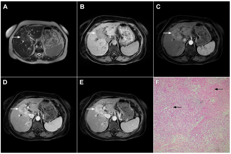
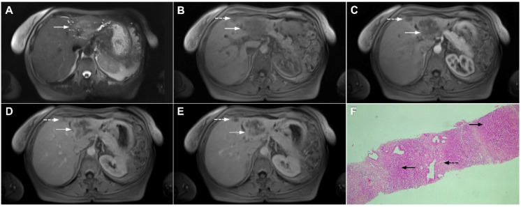
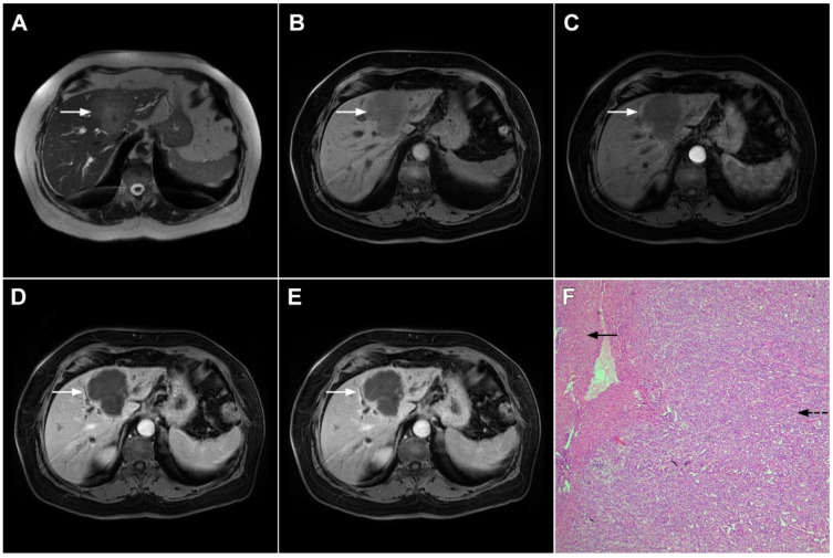
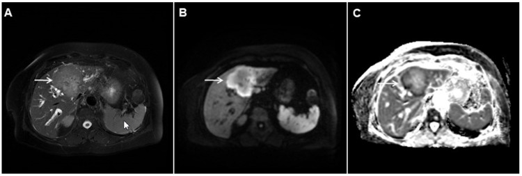
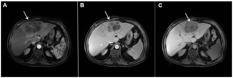
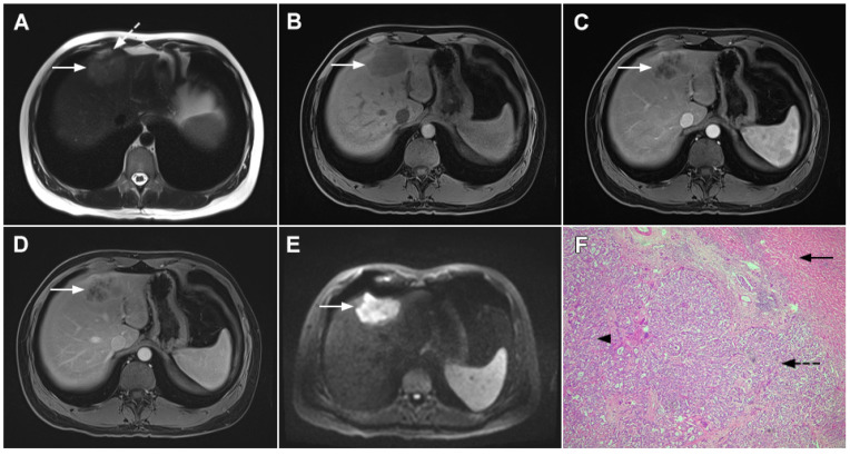
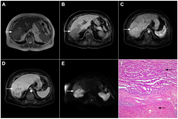
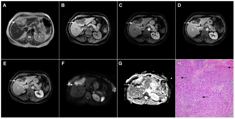
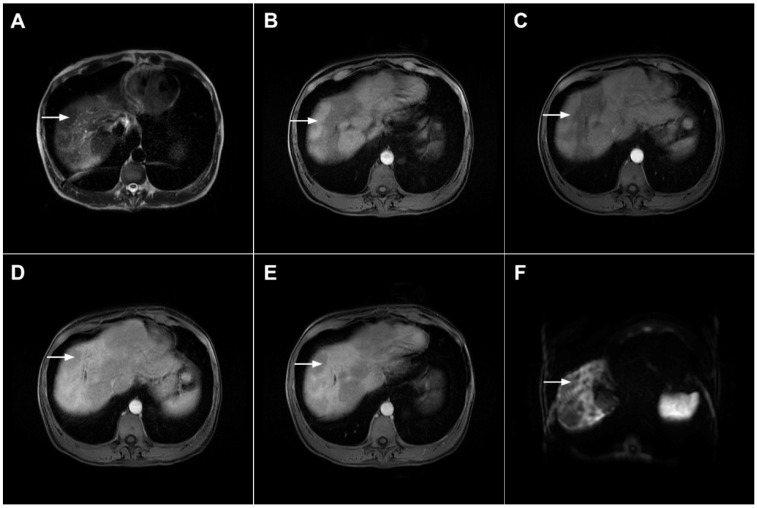
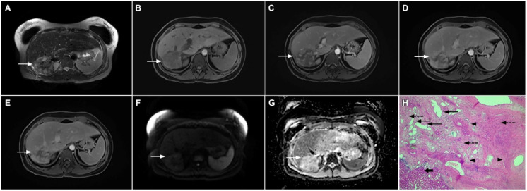
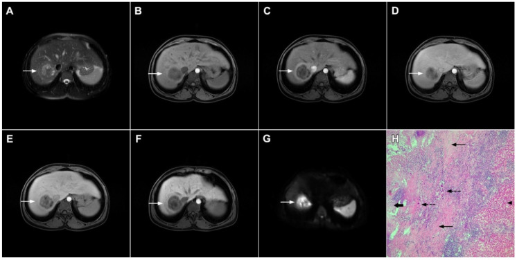
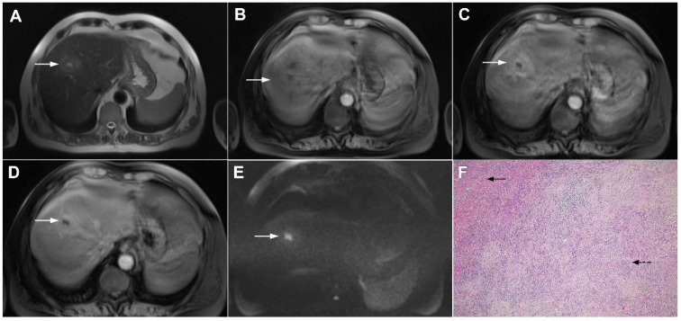
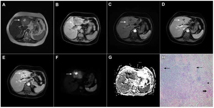
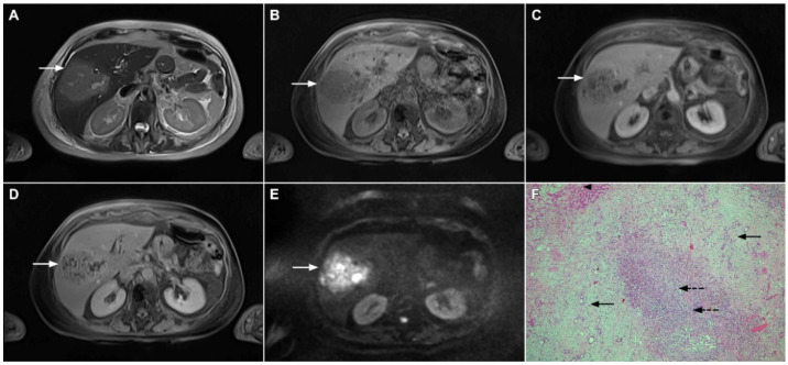
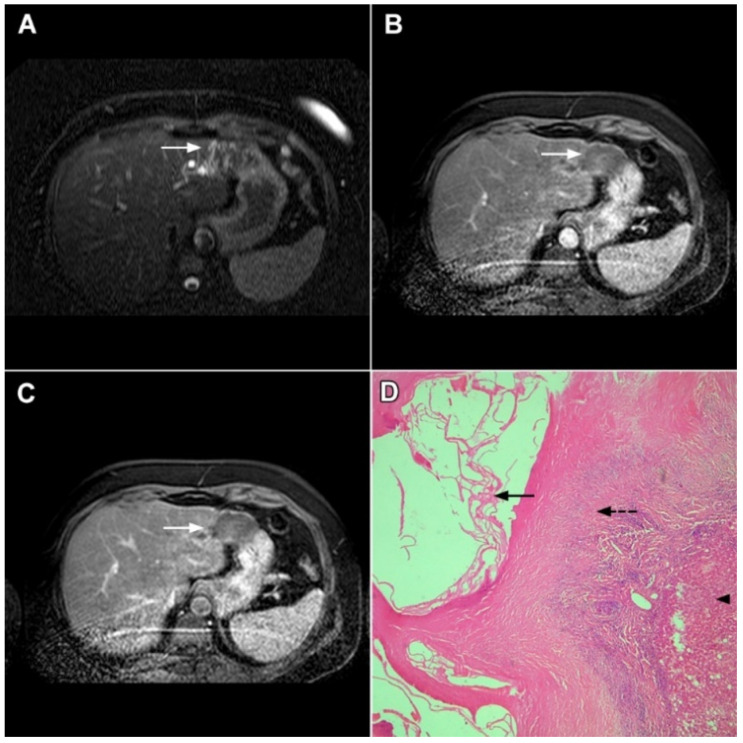
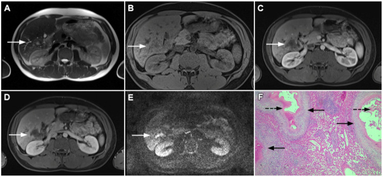
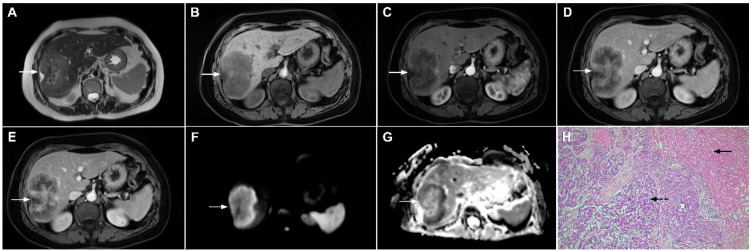
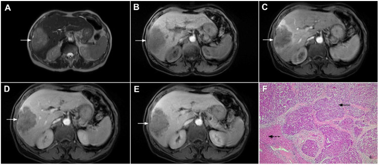
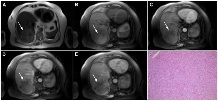
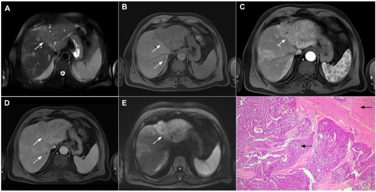
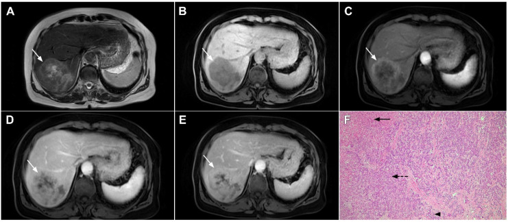
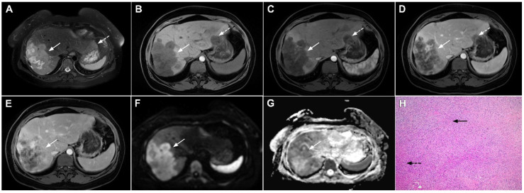
Intrahepatic cholangiocarcinoma (ICC) is the second most common primary hepatic malignancy, with mass-forming growth pattern being the most common. The typical imaging appearance of mass-forming ICC (mICC) consists of irregular ring enhancement in the arterial phase followed by the progressive central enhancement on portal venous and delayed phases. However, atypical imaging presentation in the form of hypervascular mICC might also be seen, which can be attributed to distinct pathological characteristics. Ancillary imaging features such as lobular shape, capsular retraction, segmental biliary dilatation, and vascular encasement favor the diagnosis of mICC. Nevertheless, these radiological findings may also be present in certain benign conditions such as focal confluent fibrosis, sclerosing hemangioma, organizing hepatic abscess, or the pseudosolid form of hydatid disease. In addition, a few malignant lesions including primary liver lymphoma, hemangioendothelioma, solitary hypovascular liver metastases, and atypical forms of hepatocellular carcinoma (HCC), such as scirrhous HCC, infiltrative HCC, and poorly differentiated HCC, may also pose a diagnostic dilemma by simulating mICC in imaging studies. Diffusion-weighted imaging and the use of hepatobiliary contrast agents might be helpful for differential diagnosis in certain cases. The aim of this manuscript is to provide a comprehensive overview of mICC imaging features and to describe useful tips for differential diagnosis with its potential mimickers.

摘要

肝内胆管细胞癌(ICC)是第二常见的原发性肝恶性肿瘤,以肿块型生长方式最为常见。肿块型肝内胆管细胞癌(mICC)的典型影像学表现为动脉期不规则环状强化,随后门静脉期和延迟期进行性中央强化。然而,也可能出现富血供型 mICC 的不典型表现,这可归因于其独特的病理特征。辅助影像学特征,如小叶形状、包膜回缩、节段性胆管扩张和血管包绕,有助于 mICC 的诊断。然而,这些影像学表现也可能存在于某些良性病变中,如局灶性融合性纤维增生、硬化性血管瘤、化脓性肝脓肿或包虫病的假实质性形式。此外,少数恶性病变,包括原发性肝淋巴瘤、血管内皮细胞瘤、孤立性低血供肝转移瘤和不典型的肝细胞癌(HCC)形式,如硬癌型 HCC、浸润性 HCC 和低分化 HCC,也可能在影像学研究中模拟 mICC,导致诊断困难。弥散加权成像和肝胆对比剂的使用可能有助于某些情况下的鉴别诊断。本文旨在全面概述 mICC 的影像学特征,并描述与潜在类似物进行鉴别诊断的有用技巧。

https://cdn.ncbi.nlm.nih.gov/pmc/blobs/636f/8870737/ee8f58d22050/curroncol-29-00061-g022.jpg
https://cdn.ncbi.nlm.nih.gov/pmc/blobs/636f/8870737/d44bdc3da325/curroncol-29-00061-g001.jpg
https://cdn.ncbi.nlm.nih.gov/pmc/blobs/636f/8870737/b6bda0ab3e12/curroncol-29-00061-g002.jpg
https://cdn.ncbi.nlm.nih.gov/pmc/blobs/636f/8870737/5235a240de7b/curroncol-29-00061-g003.jpg
https://cdn.ncbi.nlm.nih.gov/pmc/blobs/636f/8870737/74814188ec6d/curroncol-29-00061-g004.jpg
https://cdn.ncbi.nlm.nih.gov/pmc/blobs/636f/8870737/01a5e84d51b8/curroncol-29-00061-g005.jpg
https://cdn.ncbi.nlm.nih.gov/pmc/blobs/636f/8870737/282af450cca5/curroncol-29-00061-g006.jpg
https://cdn.ncbi.nlm.nih.gov/pmc/blobs/636f/8870737/683af707321a/curroncol-29-00061-g007.jpg
https://cdn.ncbi.nlm.nih.gov/pmc/blobs/636f/8870737/755b42c62dc6/curroncol-29-00061-g008.jpg
https://cdn.ncbi.nlm.nih.gov/pmc/blobs/636f/8870737/8e8abdf08e95/curroncol-29-00061-g009.jpg
https://cdn.ncbi.nlm.nih.gov/pmc/blobs/636f/8870737/761978e19320/curroncol-29-00061-g010.jpg
https://cdn.ncbi.nlm.nih.gov/pmc/blobs/636f/8870737/648ed2e06050/curroncol-29-00061-g011.jpg
https://cdn.ncbi.nlm.nih.gov/pmc/blobs/636f/8870737/512ebd63b278/curroncol-29-00061-g012.jpg
https://cdn.ncbi.nlm.nih.gov/pmc/blobs/636f/8870737/e198722fa4e3/curroncol-29-00061-g013.jpg
https://cdn.ncbi.nlm.nih.gov/pmc/blobs/636f/8870737/8cabb4bdd217/curroncol-29-00061-g014.jpg
https://cdn.ncbi.nlm.nih.gov/pmc/blobs/636f/8870737/170e8ab451fd/curroncol-29-00061-g015.jpg
https://cdn.ncbi.nlm.nih.gov/pmc/blobs/636f/8870737/15c8a0eca3ed/curroncol-29-00061-g016.jpg
https://cdn.ncbi.nlm.nih.gov/pmc/blobs/636f/8870737/8829fc2ef28a/curroncol-29-00061-g017.jpg
https://cdn.ncbi.nlm.nih.gov/pmc/blobs/636f/8870737/cfb5e409e623/curroncol-29-00061-g018.jpg
https://cdn.ncbi.nlm.nih.gov/pmc/blobs/636f/8870737/747b91e6c5a2/curroncol-29-00061-g019.jpg
https://cdn.ncbi.nlm.nih.gov/pmc/blobs/636f/8870737/1f46c44b10ff/curroncol-29-00061-g020.jpg
https://cdn.ncbi.nlm.nih.gov/pmc/blobs/636f/8870737/7c4459876e8f/curroncol-29-00061-g021.jpg
https://cdn.ncbi.nlm.nih.gov/pmc/blobs/636f/8870737/ee8f58d22050/curroncol-29-00061-g022.jpg

相似文献

1
Imaging Spectrum of Intrahepatic Mass-Forming Cholangiocarcinoma and Its Mimickers: How to Differentiate Them Using MRI.肝内肿块型胆管细胞癌及其模拟病变的影像学表现:MRI 如何对其进行鉴别。
Curr Oncol. 2022 Jan 30;29(2):698-723. doi: 10.3390/curroncol29020061.
2
Differentiation of mass-forming intrahepatic cholangiocarcinoma from poorly differentiated hepatocellular carcinoma: based on the multivariate analysis of contrast-enhanced computed tomography findings.基于多变量分析的增强 CT 表现对肿块型肝内胆管细胞癌与低分化肝细胞癌的鉴别诊断
Abdom Radiol (NY). 2016 May;41(5):978-89. doi: 10.1007/s00261-015-0629-z.
3
Distinguishing intrahepatic mass-forming biliary carcinomas from hepatocellular carcinoma by computed tomography and magnetic resonance imaging using the Bayesian method: a bi-center study.应用贝叶斯方法通过 CT 和 MRI 鉴别肝内肿块型胆管细胞癌和肝细胞癌:一项双中心研究。
Eur Radiol. 2020 Nov;30(11):5992-6002. doi: 10.1007/s00330-020-06972-w. Epub 2020 Jun 4.
4
Intrahepatic mass-forming cholangiocarcinoma and solitary hypovascular liver metastases: is the differential diagnosis using diffusion-weighted MRI possible?肝内肿块型胆管癌与孤立性乏血供肝转移瘤:能否通过扩散加权磁共振成像进行鉴别诊断?
Acta Radiol. 2017 Dec;58(12):1417-1426. doi: 10.1177/0284185117695666. Epub 2017 Mar 28.
5
MRI features of combined hepatocellular- cholangiocarcinoma versus mass forming intrahepatic cholangiocarcinoma.MRI 特征在肝细胞癌-胆管细胞癌与肿块型肝内胆管细胞癌的对比。
Cancer Imaging. 2018 Feb 27;18(1):8. doi: 10.1186/s40644-018-0142-z.
6
Atypical Appearance of Hepatocellular Carcinoma and Its Mimickers: How to Solve Challenging Cases Using Gadoxetic Acid-Enhanced Liver Magnetic Resonance Imaging.肝细胞癌及其类似物的不典型表现:如何使用钆塞酸增强肝脏磁共振成像解决具有挑战性的病例。
Korean J Radiol. 2019 Jul;20(7):1019-1041. doi: 10.3348/kjr.2018.0636.
7
Distinguishing intrahepatic cholangiocarcinoma from poorly differentiated hepatocellular carcinoma using precontrast and gadoxetic acid-enhanced MRI.利用造影前及钆塞酸增强磁共振成像鉴别肝内胆管癌与低分化肝细胞癌
Diagn Interv Radiol. 2015 Mar-Apr;21(2):96-104. doi: 10.5152/dir.2014.13013.
8
A convenient scoring system to distinguish intrahepatic mass-forming cholangiocarcinoma from solitary colorectal liver metastasis based on magnetic resonance imaging features.基于磁共振成像特征的一种用于鉴别肝内肿块型胆管细胞癌与单发结直肠肝转移瘤的简便评分系统。
Eur Radiol. 2023 Dec;33(12):8986-8998. doi: 10.1007/s00330-023-09873-w. Epub 2023 Jul 1.
9
Differentiation of Intrahepatic Cholangiocellular Carcinoma from Hepatocellular Carcinoma in the Cirrhotic Liver Using Contrast-enhanced MR Imaging.利用对比增强磁共振成像鉴别肝硬化肝脏中肝内胆管细胞癌与肝细胞癌
Acad Radiol. 2017 Dec;24(12):1491-1500. doi: 10.1016/j.acra.2017.06.005. Epub 2017 Jul 26.
10
Intrahepatic cholangiocarcinoma and its differential diagnosis at MRI: how radiologist should assess MR features.MRI 下肝内胆管细胞癌及其鉴别诊断:放射科医生应如何评估 MRI 特征。
Radiol Med. 2021 Dec;126(12):1584-1600. doi: 10.1007/s11547-021-01428-7. Epub 2021 Nov 29.

引用本文的文献

1
Intrahepatic cholangiocarcinoma: role of imaging as a critical component for multi-disciplinary treatment approach.肝内胆管癌:影像学作为多学科治疗方法关键组成部分的作用
Abdom Radiol (NY). 2025 Mar 17. doi: 10.1007/s00261-025-04856-5.
2
Standardizing the reporting of cholangiocarcinoma: the society of abdominal radiology disease focused panel on cholangiocarinoma lexicon.胆管癌报告的标准化:腹部放射学会胆管癌词汇疾病聚焦小组
Abdom Radiol (NY). 2025 Jul;50(7):2858-2867. doi: 10.1007/s00261-024-04769-9. Epub 2025 Jan 7.
3
Common pitfalls and diagnostic challenges in the application of LI-RADS CT/MRI algorithms: a comprehensive review.

本文引用的文献

1
Imaging features of intrahepatic cholangiocarcinoma mimicking a liver abscess: an analysis of 8 cases.肝内胆管细胞癌误诊为肝脓肿的影像学特征:8 例分析。
BMC Gastroenterol. 2021 Nov 12;21(1):427. doi: 10.1186/s12876-021-02002-1.
2
IgG4-related hepatic inflammatory pseudotumor mimicking cholangiolocellular carcinoma.IgG4 相关肝炎性假瘤,类似胆管细胞癌。
Clin J Gastroenterol. 2021 Dec;14(6):1733-1739. doi: 10.1007/s12328-021-01526-z. Epub 2021 Sep 27.
3
Liver Imaging Reporting and Data System Comprehensive Guide: MR Imaging Edition.
LI-RADS CT/MRI算法应用中的常见陷阱与诊断挑战:全面综述
Abdom Radiol (NY). 2024 Dec 24. doi: 10.1007/s00261-024-04778-8.
4
Sarcomatoid hepatocellular carcinoma with subdiaphragmatic metastasis misdiagnosed as liver abscess: A case report.伴有膈下转移的肉瘤样肝细胞癌误诊为肝脓肿:一例报告
Medicine (Baltimore). 2024 Dec 6;103(49):e40842. doi: 10.1097/MD.0000000000040842.
5
A preliminary study of developing an MRI-based model for postoperative recurrence prediction and treatment direction of intrahepatic cholangiocarcinoma.基于磁共振成像(MRI)的肝内胆管癌术后复发预测及治疗方向模型开发的初步研究。
Radiol Med. 2024 Dec;129(12):1766-1777. doi: 10.1007/s11547-024-01910-y. Epub 2024 Nov 2.
6
Prediction models for differentiating benign from malignant liver lesions based on multiparametric dual-energy non-contrast CT.基于多参数双能量非增强CT鉴别肝脏良恶性病变的预测模型
Eur Radiol. 2025 Mar;35(3):1361-1377. doi: 10.1007/s00330-024-11024-8. Epub 2024 Aug 26.
7
Tumour burden score combined with albumin-to-alkaline phosphatase ratio predicts prognosis in patients with intrahepatic cholangiocarcinoma.肿瘤负担评分联合白蛋白-碱性磷酸酶比值可预测肝内胆管细胞癌患者的预后。
J Cell Mol Med. 2024 Jul;28(13):e18530. doi: 10.1111/jcmm.18530.
8
Cholangiocarcinoma: The Current Status of Surgical Options including Liver Transplantation.胆管癌:包括肝移植在内的手术选择现状
Cancers (Basel). 2024 May 21;16(11):1946. doi: 10.3390/cancers16111946.
9
Comparison of Machine Learning Models Using Diffusion-Weighted Images for Pathological Grade of Intrahepatic Mass-Forming Cholangiocarcinoma.基于扩散加权成像的机器学习模型对肝内肿块型胆管细胞癌病理分级的比较。
J Imaging Inform Med. 2024 Oct;37(5):2252-2263. doi: 10.1007/s10278-024-01103-z. Epub 2024 Apr 16.
10
Complete regression of intrahepatic cholangiocarcinoma after right portal vein ligation. Case report.右门静脉结扎术后肝内胆管癌完全消退。病例报告。
Int J Surg Case Rep. 2024 Apr;117:109580. doi: 10.1016/j.ijscr.2024.109580. Epub 2024 Mar 24.
肝脏影像报告和数据系统综合指南:磁共振成像版。
Magn Reson Imaging Clin N Am. 2021 Aug;29(3):375-387. doi: 10.1016/j.mric.2021.05.012.
4
Intrahepatic cholangiocarcinoma mimicking a liver abscess.酷似肝脓肿的肝内胆管癌
Clin Case Rep. 2020 Aug 3;8(12):2510-2513. doi: 10.1002/ccr3.3201. eCollection 2020 Dec.
5
Futibatinib, an investigational agent for the treatment of intrahepatic cholangiocarcinoma: evidence to date and future perspectives.呋替尼,一种用于治疗肝内胆管癌的研究药物:现有证据和未来展望。
Expert Opin Investig Drugs. 2021 Apr;30(4):317-324. doi: 10.1080/13543784.2021.1837774. Epub 2020 Oct 25.
6
Differential diagnosis between hepatic alveolar echinococcosis and intrahepatic cholangiocarcinoma with conventional ultrasound and contrast-enhanced ultrasound.常规超声与超声造影对肝泡型包虫病与肝内胆管细胞癌的鉴别诊断。
BMC Med Imaging. 2020 Aug 27;20(1):101. doi: 10.1186/s12880-020-00499-8.
7
Percutaneous radiofrequency ablation in intrahepatic cholangiocarcinoma: a retrospective single-center experience.经皮射频消融治疗肝内胆管细胞癌:一项回顾性单中心经验。
Int J Hyperthermia. 2020;37(1):479-485. doi: 10.1080/02656736.2020.1763484.
8
Diagnostic approach in hepatic lymphoma: radiological imaging findings and literature review.肝淋巴瘤的诊断方法:影像学表现及文献复习。
J Cancer Res Clin Oncol. 2020 Jun;146(6):1545-1558. doi: 10.1007/s00432-020-03205-x. Epub 2020 Apr 15.
9
What happened to my liver lesion (Hepatic Sclerosed Hemangioma)? Let's not forget (radiological) history.我的肝脏病变(肝硬化性血管瘤)怎么了?咱们可别忘了(影像学)病史。
Acta Gastroenterol Belg. 2019 Oct-Dec;82(4):554-555.
10
Intrahepatic Cholangiocarcinoma.肝内胆管癌
Surg Oncol Clin N Am. 2019 Oct;28(4):587-599. doi: 10.1016/j.soc.2019.06.002.