Oral Basic Sciences, College of Dentistry, Princess Nourah Bint Abdulrahman University, Riyadh, Saudi Arabia.
Oral Biology Department, Faculty of Dentistry, Mansoura University, Mansoura, Egypt.
Stem Cell Res Ther. 2021 Nov 14;12(1):577. doi: 10.1186/s13287-021-02646-6.
Diabetes mellitus causes deterioration in the body, including serious damage of the oral cavity related to salivary gland dysfunction, characterised by hyposalivation and xerostomia. Human dental pulp stem cells (hDPSCs) represent a promising therapy source, due to the easy, minimally invasive surgical access to these cells and their high proliferative capacity. It was previously reported that the trophic support mediated by these cells can rescue the functional and structural alterations of damaged salivary glands. However, potential differentiation and paracrine effects of hDPSCs in diabetic-induced parotid gland damage have not been investigated. Our study aimed to investigate the therapeutic effects of intravenous transplantation of hDPSCs on parotid gland injury in a rat model of streptozotocin (STZ)-induced type 1 diabetes.
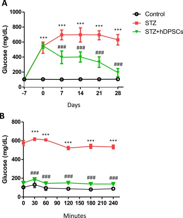
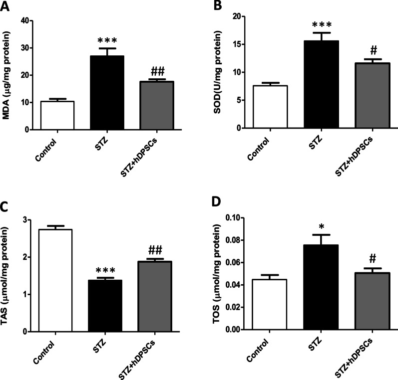
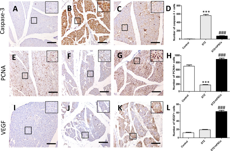
Thirty Sprague-Dawley male rats were randomly categorised into three groups: control, diabetic (STZ), and transplanted (STZ + hDPSCs). The hDPSCs or the vehicles were injected into the rats' tail veins, 7 days after STZ injection. Fasting blood glucose levels were monitored weekly. A glucose tolerance test was performed, and the parotid gland weight, salivary flow rate, oxidative stress indices, parotid gland histology, and caspase-3, vascular endothelial growth factor, proliferating cell nuclear antigen, neuronal nitric oxide synthase, endothelial nitric oxide synthase, and tetrahydrobiopterin biosynthetic enzyme expression levels in parotid tissues were assessed 28 days post-transplantation.
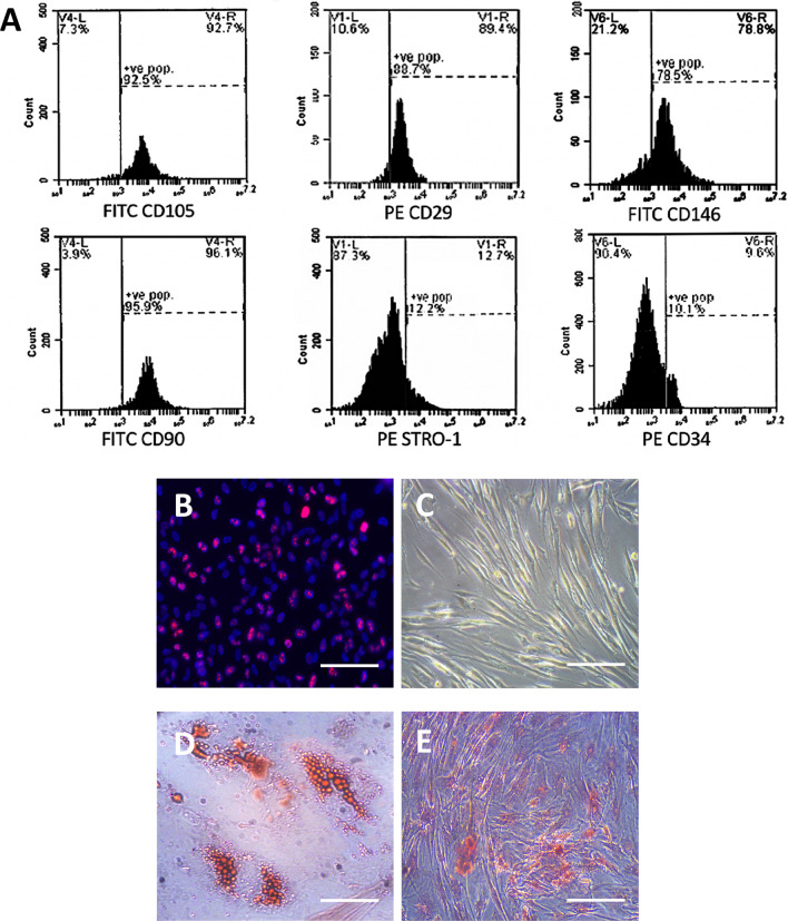
Transplantation of hDPSCs decreased blood glucose, improved parotid gland weight and salivary flow rate, and reduced oxidative stress. The cells migrated to the STZ-injured parotid gland and differentiated into acinar, ductal, and myoepithelial cells. Moreover, hDPSCs downregulated the expression of caspase-3 and upregulated the expression of vascular endothelial growth factor and proliferating cell nuclear antigen, likely exerting pro-angiogenic and anti-apoptotic effects and promoting endogenous regeneration. In addition, the transplanted cells enhanced the parotid nitric oxide-tetrahydrobiopterin pathway.
Our results showed that hDPSCs migrated to and survived within the STZ-injured parotid gland, where functional and morphological damage was prevented due to the restoration of normal glucose levels, differentiation into parotid cell populations, and stimulation of paracrine-mediated regeneration. Thus, hDPSCs may have potential in the treatment of diabetes-induced parotid gland injury.
糖尿病会导致身体恶化,包括口腔相关的唾液腺功能障碍的严重损害,其特征是唾液分泌减少和口干。人牙髓干细胞(hDPSCs)代表了一种有前途的治疗来源,因为这些细胞很容易通过微创手术获得,并且具有很高的增殖能力。先前有报道称,这些细胞的营养支持可以挽救受损唾液腺的功能和结构改变。然而,hDPSCs 在糖尿病诱导的腮腺损伤中的潜在分化和旁分泌作用尚未得到研究。我们的研究旨在研究静脉内移植 hDPSCs 对链脲佐菌素(STZ)诱导的 1 型糖尿病大鼠腮腺损伤的治疗效果。
30 只雄性 Sprague-Dawley 大鼠被随机分为三组:对照组、糖尿病组(STZ)和移植组(STZ+hDPSCs)。在 STZ 注射后 7 天,将 hDPSCs 或载体注入大鼠尾静脉。每周监测空腹血糖水平。进行葡萄糖耐量试验,并在移植后 28 天评估腮腺重量、唾液流率、氧化应激指标、腮腺组织学以及腮腺组织中 caspase-3、血管内皮生长因子、增殖细胞核抗原、神经元型一氧化氮合酶、内皮型一氧化氮合酶和四氢生物蝶呤生物合成酶的表达水平。
移植 hDPSCs 可降低血糖,改善腮腺重量和唾液流率,并减轻氧化应激。细胞迁移到 STZ 损伤的腮腺并分化为腺泡、导管和肌上皮细胞。此外,hDPSCs 下调 caspase-3 的表达,上调血管内皮生长因子和增殖细胞核抗原的表达,可能发挥促血管生成和抗细胞凋亡作用,并促进内源性再生。此外,移植细胞增强了腮腺的一氧化氮-四氢生物蝶呤途径。
我们的结果表明,hDPSCs 迁移到并存活于 STZ 损伤的腮腺中,由于正常血糖水平的恢复、向腮腺细胞群的分化以及旁分泌介导的再生刺激,防止了功能和形态损伤。因此,hDPSCs 可能在治疗糖尿病诱导的腮腺损伤方面具有潜力。