• 文献检索
  • 文档翻译
  • 深度研究
  • 学术资讯
  • Suppr Zotero 插件Zotero 插件
  • 邀请有礼
  • 套餐&价格
  • 历史记录
应用&插件
Suppr Zotero 插件Zotero 插件浏览器插件Mac 客户端Windows 客户端微信小程序
定价
高级版会员购买积分包购买API积分包
服务
文献检索文档翻译深度研究API 文档MCP 服务
关于我们
关于 Suppr公司介绍联系我们用户协议隐私条款
关注我们

Suppr 超能文献

核心技术专利:CN118964589B侵权必究
粤ICP备2023148730 号-1Suppr @ 2026

文献检索

告别复杂PubMed语法,用中文像聊天一样搜索,搜遍4000万医学文献。AI智能推荐,让科研检索更轻松。

立即免费搜索

文件翻译

保留排版,准确专业,支持PDF/Word/PPT等文件格式,支持 12+语言互译。

免费翻译文档

深度研究

AI帮你快速写综述,25分钟生成高质量综述,智能提取关键信息,辅助科研写作。

立即免费体验

受刺激的米勒细胞的蛋白质组学表型分析揭示了强烈的促炎信号传导和抗原呈递能力。

Proteomic Phenotyping of Stimulated Müller Cells Uncovers Profound Pro-Inflammatory Signaling and Antigen-Presenting Capacity.

作者信息

Schmalen Adrian, Lorenz Lea, Grosche Antje, Pauly Diana, Deeg Cornelia A, Hauck Stefanie M

机构信息

Research Unit Protein Science and Metabolomics and Proteomics Core, Helmholtz Center Munich, German Research Center for Environmental Health (GmbH), Neuherberg, Germany.

Chair of Physiology, Department of Veterinary Sciences, LMU Munich, Martinsried, Germany.

出版信息

Front Pharmacol. 2021 Oct 29;12:771571. doi: 10.3389/fphar.2021.771571. eCollection 2021.

DOI:10.3389/fphar.2021.771571
PMID:34776983
原文链接:https://pmc.ncbi.nlm.nih.gov/articles/PMC8585775/
Abstract

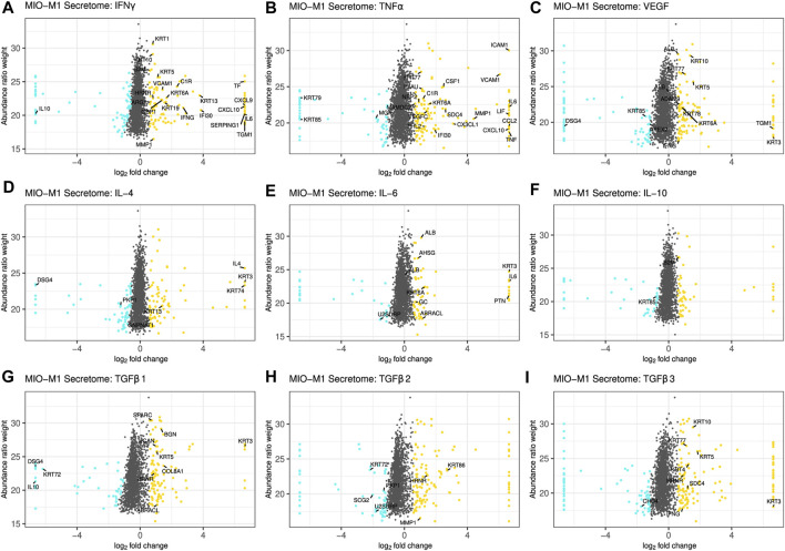
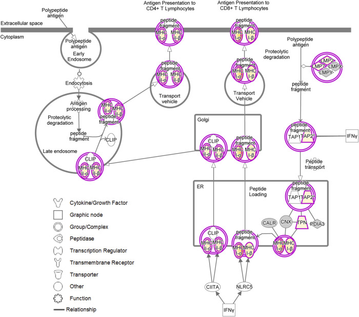
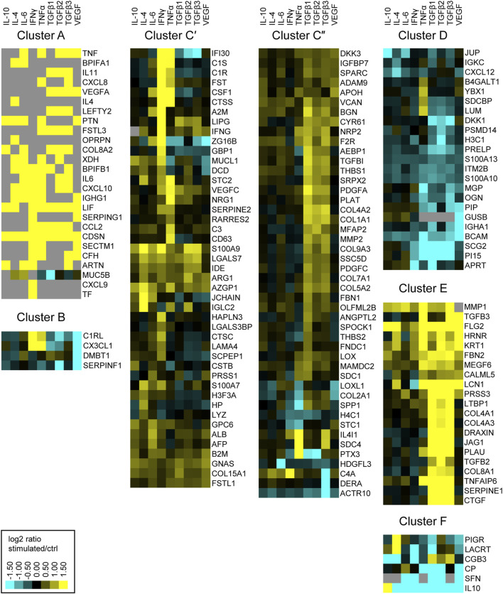
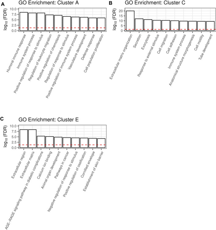
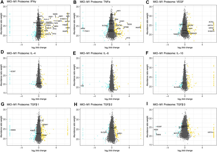
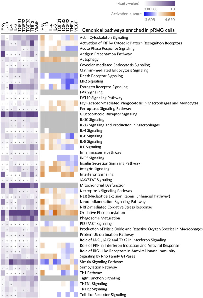
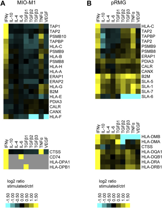
Müller cells are the main macroglial cells of the retina exerting a wealth of functions to maintain retinal homoeostasis. Upon pathological changes in the retina, they become gliotic with both protective and detrimental consequences. Accumulating data also provide evidence for a pivotal role of Müller cells in the pathogenesis of diabetic retinopathy (DR). While microglial cells, the resident immune cells of the retina are considered as main players in inflammatory processes associated with DR, the implication of activated Müller cells in chronic retinal inflammation remains to be elucidated. In order to assess the signaling capacity of Müller cells and their role in retinal inflammation, we performed in-depth proteomic analysis of Müller cell proteomes and secretomes after stimulation with INFγ, TNFα, IL-4, IL-6, IL-10, VEGF, TGFβ1, TGFβ2 and TGFβ3. We used both, primary porcine Müller cells and the human Müller cell line MIO-M1 for our hypothesis generating approach. Our results point towards an intense signaling capacity of Müller cells, which reacted in a highly discriminating manner upon treatment with different cytokines. Stimulation of Müller cells resulted in a primarily pro-inflammatory phenotype with secretion of cytokines and components of the complement system. Furthermore, we observed evidence for mitochondrial dysfunction, implying oxidative stress after treatment with the various cytokines. Finally, both MIO-M1 cells and primary porcine Müller cells showed several characteristics of atypical antigen-presenting cells, as they are capable of inducing MHC class I and MHC class II with co-stimulatory molecules. In line with this, they express proteins associated with formation and maturation of phagosomes. Thus, our findings underline the importance of Müller cell signaling in the inflamed retina, indicating an active role in chronic retinal inflammation.

摘要

缪勒细胞是视网膜主要的大胶质细胞,发挥着众多维持视网膜稳态的功能。视网膜发生病理变化时,它们会发生胶质化,产生保护和有害两种后果。越来越多的数据也证明缪勒细胞在糖尿病视网膜病变(DR)发病机制中起关键作用。虽然视网膜常驻免疫细胞小胶质细胞被认为是与DR相关炎症过程的主要参与者,但活化的缪勒细胞在慢性视网膜炎症中的作用仍有待阐明。为了评估缪勒细胞的信号传导能力及其在视网膜炎症中的作用,我们在用INFγ、TNFα、IL-4、IL-6、IL-10、VEGF、TGFβ1、TGFβ2和TGFβ3刺激后,对缪勒细胞蛋白质组和分泌蛋白质组进行了深入的蛋白质组学分析。我们使用原代猪缪勒细胞和人缪勒细胞系MIO-M1进行我们的假设生成方法。我们的结果表明缪勒细胞具有强大的信号传导能力,在用不同细胞因子处理时会以高度有区别的方式做出反应。缪勒细胞的刺激导致主要的促炎表型,伴有细胞因子和补体系统成分的分泌。此外,我们观察到线粒体功能障碍的证据,这意味着在用各种细胞因子处理后存在氧化应激。最后,MIO-M1细胞和原代猪缪勒细胞都表现出非典型抗原呈递细胞的几个特征,因为它们能够诱导带有共刺激分子的MHC I类和MHC II类。与此一致的是,它们表达与吞噬体形成和成熟相关的蛋白质。因此,我们的发现强调了缪勒细胞信号传导在炎症视网膜中的重要性,表明其在慢性视网膜炎症中发挥积极作用。

https://cdn.ncbi.nlm.nih.gov/pmc/blobs/31b7/8585775/4700f4eb1599/fphar-12-771571-g007.jpg
https://cdn.ncbi.nlm.nih.gov/pmc/blobs/31b7/8585775/ea3729a895e6/fphar-12-771571-g001.jpg
https://cdn.ncbi.nlm.nih.gov/pmc/blobs/31b7/8585775/cb22b6016164/fphar-12-771571-g002.jpg
https://cdn.ncbi.nlm.nih.gov/pmc/blobs/31b7/8585775/4cc1e71d8f18/fphar-12-771571-g003.jpg
https://cdn.ncbi.nlm.nih.gov/pmc/blobs/31b7/8585775/68f80b18c234/fphar-12-771571-g004.jpg
https://cdn.ncbi.nlm.nih.gov/pmc/blobs/31b7/8585775/0484a9d3ffaf/fphar-12-771571-g005.jpg
https://cdn.ncbi.nlm.nih.gov/pmc/blobs/31b7/8585775/619b18641b6d/fphar-12-771571-g006.jpg
https://cdn.ncbi.nlm.nih.gov/pmc/blobs/31b7/8585775/4700f4eb1599/fphar-12-771571-g007.jpg

相似文献

1
Proteomic Phenotyping of Stimulated Müller Cells Uncovers Profound Pro-Inflammatory Signaling and Antigen-Presenting Capacity.受刺激的米勒细胞的蛋白质组学表型分析揭示了强烈的促炎信号传导和抗原呈递能力。
Front Pharmacol. 2021 Oct 29;12:771571. doi: 10.3389/fphar.2021.771571. eCollection 2021.
2
Müller glia as an important source of cytokines and inflammatory factors present in the gliotic retina during proliferative vitreoretinopathy.缪勒胶质细胞是增殖性玻璃体视网膜病变期间胶质化视网膜中细胞因子和炎症因子的重要来源。
Glia. 2016 Apr;64(4):495-506. doi: 10.1002/glia.22942. Epub 2015 Nov 10.
3
Endothelin B Receptors on Primary Chicken Müller Cells and the Human MIO-M1 Müller Cell Line Activate ERK Signaling via Transactivation of Epidermal Growth Factor Receptors.原代鸡 Müller 细胞和人 MIO-M1 Müller 细胞系上的内皮素 B 受体通过表皮生长因子受体的反式激活激活 ERK 信号通路。
PLoS One. 2016 Dec 8;11(12):e0167778. doi: 10.1371/journal.pone.0167778. eCollection 2016.
4
Müller cells and diabetic retinopathy.米勒细胞与糖尿病视网膜病变
Vision Res. 2017 Oct;139:93-100. doi: 10.1016/j.visres.2017.03.013. Epub 2017 Sep 5.
5
Basic fibroblast growth factor contributes to a shift in the angioregulatory activity of retinal glial (Müller) cells.碱性成纤维细胞生长因子有助于改变视网膜神经胶质(Müller)细胞的血管生成调节活性。
PLoS One. 2013 Jul 4;8(7):e68773. doi: 10.1371/journal.pone.0068773. Print 2013.
6
Loss of X-box binding protein 1 in Müller cells augments retinal inflammation in a mouse model of diabetes.Müller 细胞中 X 盒结合蛋白 1 的缺失可增强糖尿病小鼠模型的视网膜炎症。
Diabetologia. 2019 Mar;62(3):531-543. doi: 10.1007/s00125-018-4776-y. Epub 2019 Jan 6.
7
Bone morphogenetic protein 2: a potential new player in the pathogenesis of diabetic retinopathy.骨形态发生蛋白 2:糖尿病视网膜病变发病机制中的一个潜在新角色。
Exp Eye Res. 2014 Aug;125:79-88. doi: 10.1016/j.exer.2014.05.012. Epub 2014 Jun 6.
8
Plasma Rich in Growth Factors (PRGF) Increases the Number of Retinal Müller Glia in Culture but Not the Survival of Retinal Neurons.富含生长因子的血浆(PRGF)可增加培养的视网膜穆勒胶质细胞数量,但对视网膜神经元的存活无影响。
Front Pharmacol. 2021 Mar 9;12:606275. doi: 10.3389/fphar.2021.606275. eCollection 2021.
9
Cell Surface Profiling of Retinal Müller Glial Cells Reveals Association to Immune Pathways after LPS Stimulation.视网膜神经胶质细胞表面分析显示 LPS 刺激后与免疫途径相关。
Cells. 2021 Mar 23;10(3):711. doi: 10.3390/cells10030711.
10
Rho-Associated Protein Kinase-1 Mediates the Regulation of Inflammatory Markers in Diabetic Retina and in Retinal Müller Cells.Rho相关蛋白激酶-1介导糖尿病视网膜及视网膜Müller细胞中炎症标志物的调控。
Ann Clin Lab Sci. 2018 Mar;48(2):137-145.

引用本文的文献

1
Interferons and shape PD-L1 regulation in retinal barrier cells: the critical role of proteases.干扰素与视网膜屏障细胞中PD-L1的调控:蛋白酶的关键作用。
Front Immunol. 2025 Jun 17;16:1607247. doi: 10.3389/fimmu.2025.1607247. eCollection 2025.
2
Differential Expression of ARG1 and MRC2 in Retinal Müller Glial Cells During Autoimmune Uveitis.自身免疫性葡萄膜炎期间视网膜Müller胶质细胞中ARG1和MRC2的差异表达
Biomolecules. 2025 Feb 14;15(2):288. doi: 10.3390/biom15020288.
3
Glia Modulates Immune Responses in the Retina Through Distinct MHC Pathways.

本文引用的文献

1
High glucose treatment promotes extracellular matrix proteome remodeling in Mller glial cells.高糖处理促进穆勒胶质细胞的细胞外基质蛋白质组重塑。
PeerJ. 2021 May 18;9:e11316. doi: 10.7717/peerj.11316. eCollection 2021.
2
Molecular Signatures of Idiopathic Pulmonary Fibrosis.特发性肺纤维化的分子特征。
Am J Respir Cell Mol Biol. 2021 Oct;65(4):430-441. doi: 10.1165/rcmb.2020-0546OC.
3
TNF-α released from retinal Müller cells aggravates retinal pigment epithelium cell apoptosis by upregulating mitophagy during diabetic retinopathy.
神经胶质细胞通过不同的主要组织相容性复合体途径调节视网膜中的免疫反应。
Glia. 2025 Apr;73(4):822-839. doi: 10.1002/glia.24656. Epub 2025 Jan 28.
4
Candidate SNP Markers Significantly Altering the Affinity of the TATA-Binding Protein for the Promoters of Human Genes Associated with Primary Open-Angle Glaucoma.候选单核苷酸多态性标记显著改变TATA结合蛋白与原发性开角型青光眼相关人类基因启动子的亲和力。
Int J Mol Sci. 2024 Nov 28;25(23):12802. doi: 10.3390/ijms252312802.
5
Induction, amplification, and propagation of diabetic retinopathy-associated inflammatory cytokines between human retinal microvascular endothelial and Müller cells and in the mouse retina.诱导、放大和传播人视网膜微血管内皮细胞和 Muller 细胞以及小鼠视网膜中与糖尿病视网膜病变相关的炎症细胞因子。
Cell Signal. 2024 Dec;124:111454. doi: 10.1016/j.cellsig.2024.111454. Epub 2024 Oct 9.
6
Photoreceptor regeneration occurs normally in microglia-deficient irf8 mutant zebrafish following acute retinal damage.急性视网膜损伤后,在小胶质细胞缺陷型 irf8 突变斑马鱼中视细胞能够正常再生。
Sci Rep. 2024 Aug 30;14(1):20146. doi: 10.1038/s41598-024-70859-9.
7
Molecular mechanisms underlying inherited photoreceptor degeneration as targets for therapeutic intervention.作为治疗干预靶点的遗传性光感受器变性的分子机制。
Front Cell Neurosci. 2024 Feb 2;18:1343544. doi: 10.3389/fncel.2024.1343544. eCollection 2024.
8
The glucocorticoid receptor as a master regulator of the Müller cell response to diabetic conditions in mice.糖皮质激素受体作为小鼠 Müller 细胞对糖尿病状态反应的主要调节因子。
J Neuroinflammation. 2024 Jan 25;21(1):33. doi: 10.1186/s12974-024-03021-x.
9
NMOSD IgG Impact Retinal Cells in Murine Retinal Explants.视神经脊髓炎谱系障碍免疫球蛋白G影响小鼠视网膜外植体中的视网膜细胞。
Curr Issues Mol Biol. 2023 Sep 7;45(9):7319-7335. doi: 10.3390/cimb45090463.
10
Different responses of the MIO‑M1 Mueller cell line to angiotensin II under hyperglycemic or hypoxic conditions.在高血糖或缺氧条件下,MIO-M1穆勒细胞系对血管紧张素II的不同反应。
Biomed Rep. 2023 Aug 7;19(3):62. doi: 10.3892/br.2023.1644. eCollection 2023 Sep.
糖尿病视网膜病变中,视网膜 Müller 细胞释放的 TNF-α 通过上调细胞自噬加剧了视网膜色素上皮细胞的凋亡。
Biochem Biophys Res Commun. 2021 Jul 5;561:143-150. doi: 10.1016/j.bbrc.2021.05.027. Epub 2021 May 21.
4
Cell Surface Profiling of Retinal Müller Glial Cells Reveals Association to Immune Pathways after LPS Stimulation.视网膜神经胶质细胞表面分析显示 LPS 刺激后与免疫途径相关。
Cells. 2021 Mar 23;10(3):711. doi: 10.3390/cells10030711.
5
NEU1 is more abundant in uveitic retina with concomitant desialylation of retinal cells.NEU1在葡萄膜炎性视网膜中更为丰富,同时视网膜细胞会发生去唾液酸化。
Glycobiology. 2021 Aug 7;31(7):873-883. doi: 10.1093/glycob/cwab014.
6
Retinal Inflammation, Cell Death and Inherited Retinal Dystrophies.视网膜炎症、细胞死亡与遗传性视网膜变性。
Int J Mol Sci. 2021 Feb 20;22(4):2096. doi: 10.3390/ijms22042096.
7
Causes of blindness and vision impairment in 2020 and trends over 30 years, and prevalence of avoidable blindness in relation to VISION 2020: the Right to Sight: an analysis for the Global Burden of Disease Study.2020 年失明和视力障碍的原因及 30 多年来的趋势,以及与 VISION 2020:看见的权利相关的可避免盲的患病率:全球疾病负担研究的分析。
Lancet Glob Health. 2021 Feb;9(2):e144-e160. doi: 10.1016/S2214-109X(20)30489-7. Epub 2020 Dec 1.
8
The Role of Inflammation in Diabetic Retinopathy.炎症在糖尿病视网膜病变中的作用。
Front Immunol. 2020 Nov 6;11:583687. doi: 10.3389/fimmu.2020.583687. eCollection 2020.
9
Microglia and Inflammatory Responses in Diabetic Retinopathy.小胶质细胞与糖尿病视网膜病变中的炎症反应。
Front Immunol. 2020 Nov 6;11:564077. doi: 10.3389/fimmu.2020.564077. eCollection 2020.
10
Inflammation and diabetic retinopathy.炎症与糖尿病视网膜病变。
Mol Vis. 2020 Oct 15;26:718-721. eCollection 2020.