Beuse Ansgar, Deissler Heidrun L, Hollborn Margrit, Unterlauft Jan Darius, Busch Catharina, Rehak Matus
Department of Ophthalmology, University of Leipzig, D-04103 Leipzig, Germany.
Department of Ophthalmology, Justus-Liebig-University Giessen, D-35392 Giessen, Germany.
Biomed Rep. 2023 Aug 7;19(3):62. doi: 10.3892/br.2023.1644. eCollection 2023 Sep.
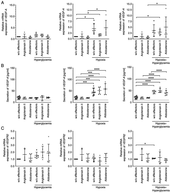
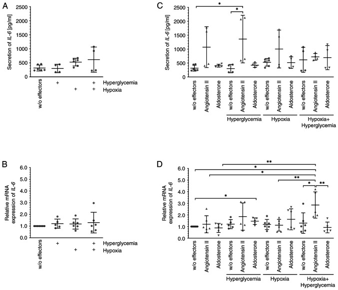
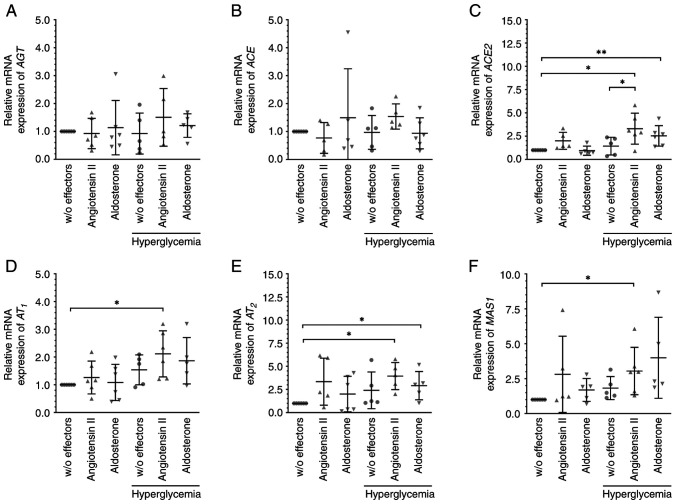
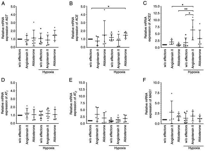
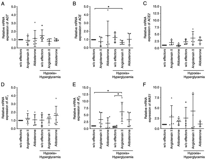
Members of the renin-angiotensin aldosterone system (RAAS) are expressed by various retinal tissues including Mueller glial cells. As the RAAS is hypothesized to play an important role in the pathogenesis of diseases that threaten vision, such as diabetic macular edema or retinal vein occlusion, the possible changes induced by exposure of the human cell line MIO-M1, an established model of Mueller cells, to angiotensin II or aldosterone for 6 h under hypoxic and/or hyperglycemic conditions were investigated. The mRNA expression levels of the members of the RAAS were assessed by reverse transcription-quantitative PCR, and the secretion of cytokines was assessed by ELISA. Under hyperglycemic conditions, the mRNA expression levels of the angiotensin-converting enzyme 2 (ACE2), angiotensin II receptors, AT and AT, and the receptor of angiotensin (1-7) MAS1 were significantly higher after exposure to angiotensin II, and the expression of ACE2, AT, and IL-6 (a marker of inflammation) was significantly increased after treatment with aldosterone; the expression of the other targets investigated remained unchanged. Significantly more IL-6 was secreted by MIO-M1 cells exposed to hyperglycemia and angiotensin. When cells were cultured in a hypoxic environment, additional treatment with aldosterone significantly increased the mRNA expression levels of ACE, but significantly more ACE2 mRNA was expressed in the presence of angiotensin II. Under hypoxic plus hyperglycemic conditions, significantly less ACE but more AT was expressed after treatment with angiotensin II, which also led to strongly elevated expression of IL-6. The mRNA expression levels of the angiogenic growth factor VEGF-A and secretion of the encoded protein were notably increased under hypoxic and hypoxic plus hyperglycemic conditions, irrespective of additional treatment with angiotensin II or aldosterone. These findings suggest that angiotensin II induces a pro-inflammatory response in MIO-M1 cells under hyperglycemic conditions despite activation of the counteracting ACE2/MAS1 signaling cascade. However, hypoxia results in an increased expression of angiogenic VEGF-A by these cells, which is not altered by angiotensin II or aldosterone.
肾素-血管紧张素-醛固酮系统(RAAS)的成员由包括穆勒胶质细胞在内的各种视网膜组织表达。由于RAAS被认为在威胁视力的疾病(如糖尿病性黄斑水肿或视网膜静脉阻塞)的发病机制中起重要作用,因此研究了在缺氧和/或高血糖条件下,将人细胞系MIO-M1(一种已建立的穆勒细胞模型)暴露于血管紧张素II或醛固酮6小时所诱导的可能变化。通过逆转录定量PCR评估RAAS成员的mRNA表达水平,并通过酶联免疫吸附测定法评估细胞因子的分泌。在高血糖条件下,暴露于血管紧张素II后,血管紧张素转换酶2(ACE2)、血管紧张素II受体AT1和AT2以及血管紧张素(1-7)受体MAS1的mRNA表达水平显著升高,用醛固酮处理后,ACE2、AT1和白细胞介素-6(炎症标志物)的表达显著增加;所研究的其他靶点的表达保持不变。暴露于高血糖和血管紧张素的MIO-M1细胞分泌的白细胞介素-6明显更多。当细胞在缺氧环境中培养时,额外用醛固酮处理显著增加了ACE的mRNA表达水平,但在存在血管紧张素II的情况下,ACE2 mRNA的表达明显更多。在缺氧加高血糖条件下,用血管紧张素II处理后,ACE的表达明显减少,但AT1的表达增加,这也导致白细胞介素-6的表达强烈升高。无论是否额外用血管紧张素II或醛固酮处理,在缺氧和缺氧加高血糖条件下,血管生成生长因子VEGF-A的mRNA表达水平和编码蛋白的分泌都显著增加。这些发现表明,尽管激活了起抵消作用的ACE2/MAS1信号级联反应,但在高血糖条件下,血管紧张素II仍会在MIO-M1细胞中诱导促炎反应。然而,缺氧会导致这些细胞中血管生成性VEGF-A的表达增加,而血管紧张素II或醛固酮不会改变这种情况。