Department of Endodontics, Faculty of Dentistry, King Abdulaziz University, Jeddah, Saudi Arabia.
Department of Dental Public Health, Faculty of Dentistry, King Abdulaziz University, Jeddah, Saudi Arabia.
BMC Oral Health. 2021 Nov 19;21(1):595. doi: 10.1186/s12903-021-01961-x.
Various systems of nickel-titanium (NiTi) instrument have long been commercially available. However, the preparation of narrow and curved root canals has always been challenging. The purpose of this study was to compare the shaping ability of two NiTi systems (2Shape and NeoNiTi) in severely curved root canals with different morphological patterns using micro-computed tomography (Micro-CT).
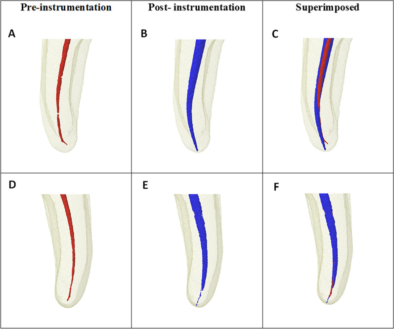
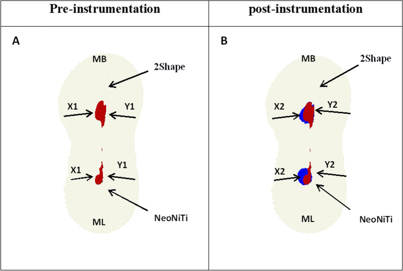
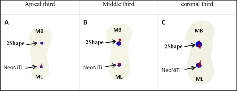
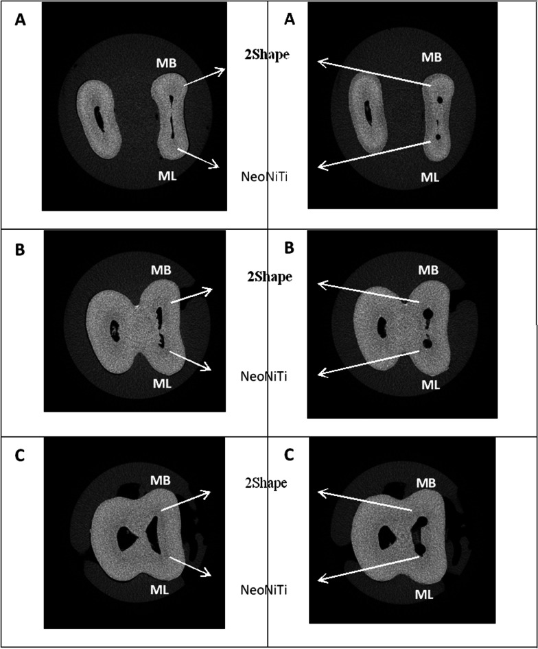
A total of 22 human extracted permanent teeth of mandibular first molars, with the exact mesial angle of curvature of 25 and 35 degrees, according to Schneider's technique, were distributed randomly into two groups (group I: 2Shape, group II: NeoNiTi) based on the rotary system used (n = 22). The groups were subdivided into two subgroups corresponding to the angle of canal curvature (25° and 35°) (n = 11). Canals were scanned using Micro-CT pre- and post-preparation to assess the volume of dentin removed; canal transportation; and canal centering ratio at 3, 6, and 9 mm from the apex. The Mann-Whitney U test was utilized to determine any significant differences between the two systems. The level of statistical significance was set at p < 0.05.
There was no significant difference between the two groups in volume of dentin removed; canal transportation; and centering ability for 25° and 35° canal curvatures at 3, 6, and 9 mm from the apex (coronal, middle, and apical) thirds (p > 0.05). At the middle third, the NeoNiTi group demonstrated a statistically significant increase in volume of dentin removed for 35° canal curvatures compared to the 2Shape group.
Within the limitation of our in vitro study, 2Shape and NeoNiTi systems with severely curved canals were confirmed to be relatively safe in preparation and to respect original canal anatomy. Nevertheless, NeoNiTi instruments produced more centered preparation and minimal canal deviation compared to the 2Shape system.
各种镍钛(NiTi)器械系统早已在商业上广泛应用。然而,预备狭窄且弯曲的根管一直具有挑战性。本研究旨在使用微计算机断层扫描(Micro-CT)比较两种 NiTi 系统(2Shape 和 NeoNiTi)在不同形态弯曲根管中的成形能力。
根据 Schneider 技术,将 22 颗下颌第一磨牙的人离体恒牙随机分为两组(组 I:2Shape,组 II:NeoNiTi),根据使用的旋转系统(n=22)。根据根管弯曲角度(25°和 35°)(n=11)将两组再分为两个亚组。在预备前后使用 Micro-CT 扫描根管,以评估牙本质去除量;根管偏移;以及距根尖 3、6 和 9mm 处的根管中心率。使用曼-惠特尼 U 检验比较两种系统之间是否存在显著差异。统计学显著性水平设定为 p<0.05。
在去除牙本质量、根管偏移和 3、6 和 9mm 处(冠部、中部和根尖部)的根管中心能力方面,两组之间在 25°和 35°根管弯曲角度之间无显著差异(p>0.05)。在中部,与 2Shape 组相比,NeoNiTi 组在 35°根管弯曲角度下牙本质去除量有统计学显著增加。
在我们的体外研究的限制范围内,对于严重弯曲的根管,2Shape 和 NeoNiTi 系统被证实是相对安全的,并且可以保留原始根管解剖结构。然而,与 2Shape 系统相比,NeoNiTi 器械的制备更居中,根管偏移更小。