Zhang Jie, Gao Feng, Ma Yuqian, Xue Tian, Shen Yong
Institute on Aging and Brain Disorders, The First Affiliated Hospital of USTC, Hefei National Laboratory for Physical Sciences at the Microscale, School of Life Science, Division of Life Science and Medicine, University of Science and Technology of China, Hefei 230026, China.
Neurodegenerative Disorder Research Center, CAS Key Laboratory of Brain Function and Disease, University of Science and Technology of China, Hefei 230026, China.
iScience. 2021 Oct 21;24(11):103327. doi: 10.1016/j.isci.2021.103327. eCollection 2021 Nov 19.
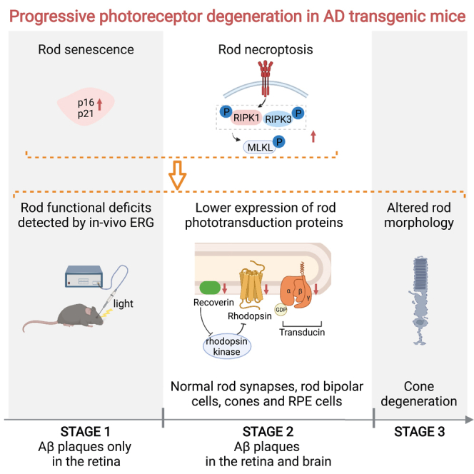
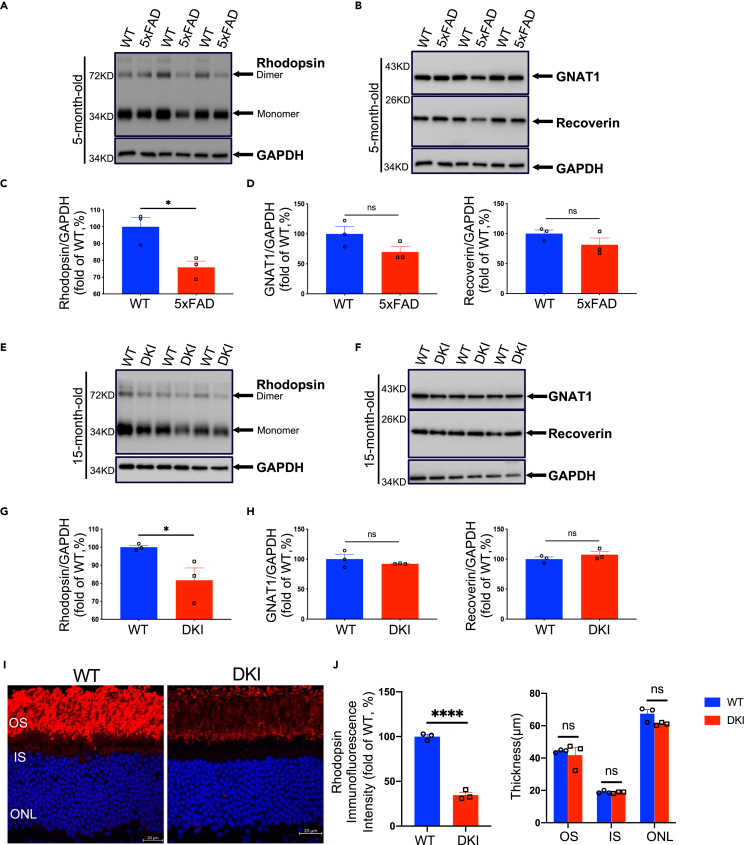
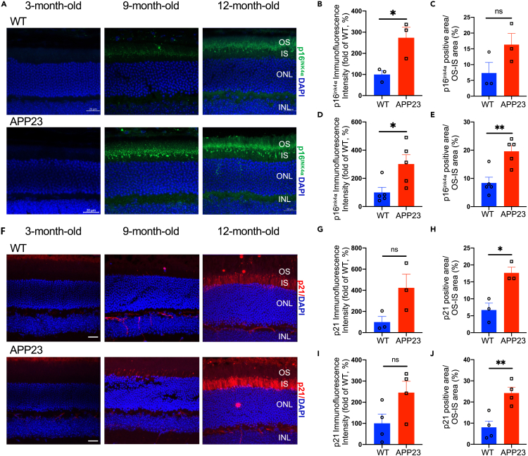
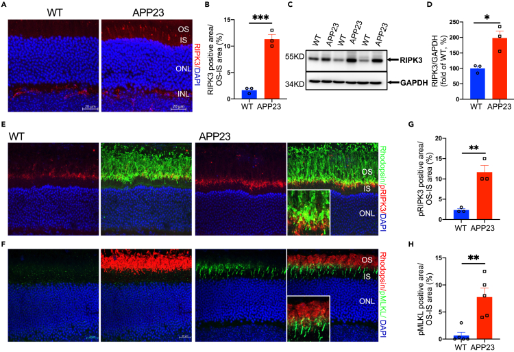
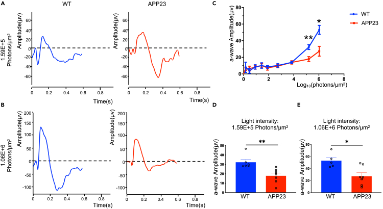
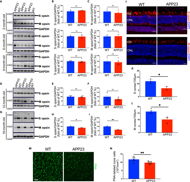
Light sensitivity of the vertebrate retina relies on the integrity of photoreceptors, including rods and cones. Research in patients with Alzheimer's disease (AD) and in AD transgenic mice reports that accumulated amyloid-β (Aβ) plaques in the retina are toxic to retinal neurons. Moreover, Aβ plaques are deposited around the rods and cones, yet photoreceptor anomalies remain unclear in AD. Here, we identify the progressive degeneration of rods and cones characterized by impaired expression of phototransduction proteins, morphological alterations, functional deficits, and even cell loss. Furthermore, we demonstrate that cell senescence and necroptosis were involved in rod degeneration. Importantly, using in vivo scotopic electroretinogram, we detected rod degeneration in early-stage AD transgenic mice before Aβ plaques were observed in the brain. Moreover, we demonstrate that rod degeneration was among the earliest AD retinal manifestations compared with other types of retinal neurons. Overall, our study is the first to identify and detect in vivo, early-onset photoreceptor degeneration in AD.
脊椎动物视网膜的光敏感性依赖于光感受器(包括视杆细胞和视锥细胞)的完整性。对阿尔茨海默病(AD)患者和AD转基因小鼠的研究报告称,视网膜中积累的淀粉样β蛋白(Aβ)斑块对视网膜神经元有毒性。此外,Aβ斑块沉积在视杆细胞和视锥细胞周围,但AD中光感受器异常仍不清楚。在这里,我们确定了视杆细胞和视锥细胞的进行性退化,其特征为光转导蛋白表达受损、形态改变、功能缺陷,甚至细胞丢失。此外,我们证明细胞衰老和坏死性凋亡参与了视杆细胞退化。重要的是,使用体内暗视视网膜电图,我们在脑内观察到Aβ斑块之前,在早期AD转基因小鼠中检测到了视杆细胞退化。此外,我们证明与其他类型的视网膜神经元相比,视杆细胞退化是AD最早的视网膜表现之一。总体而言,我们的研究首次在体内鉴定并检测到AD中早发性光感受器退化。