Acibadem University, Department of Medical Imaging, Istanbul, Turkey.
University of Iowa, Hospital and Clinics, Department of Radiology, 200 Hawkins Drive, Iowa City, IA 52242.
Acad Radiol. 2022 Jan;29(1):31-41. doi: 10.1016/j.acra.2021.10.019. Epub 2021 Oct 27.
To evaluate how COVID-19 anosmia imaging findings resembled and differed from postinfectious olfactory dysfunction (OD).
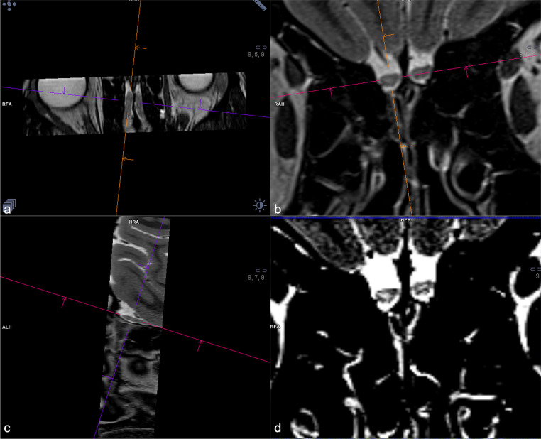
A total of 31 patients presenting with persistent COVID-19 related OD and 97 patients with post-infectious OD were included. Olfactory bulb MRI, DTI and olfactory fMRI findings in both groups were retrospectively assessed.
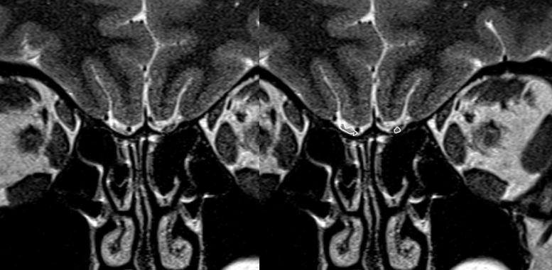
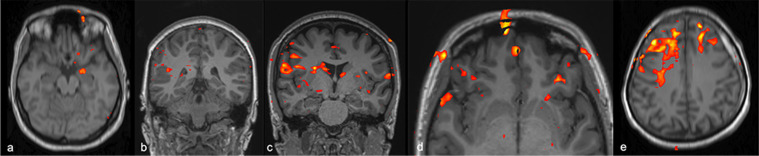
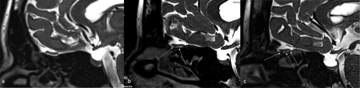
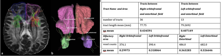
All COVID-19 related OD cases were anosmic, 18.6% of post-infectious OD patients were hyposmic and remaining 81.4% were anosmic. Mean interval between onset of OD and imaging was 1.5 months for COVID-19 related OD and 6 months for post-infectious OD. Olfactory bulb volumes were significantly higher in COVID-19 related OD than post-infectious OD. Deformed bulb morphology and increased olfactory bulb signal intensity was seen in 58.1% and 51.6% with COVID-19 related OD; and 63.9% - 46.4% with post-infectious OD; without significant difference. Significantly higher rate of olfactory nerve clumping and higher QA values at orbitofrontal and entorhinal regions were observed in COVID-19 related OD than post-infectious OD. Absence of orbitofrontal and entorhinal activity showed no statistically significant difference between COVID-19 related OD and post-infectious OD, however trigeminosensory activity was more robust in COVID-19 related OD cases.
Olfactory bulb damage may play a central role in persistent COVID-19 related anosmia. Though there is decreased olfactory bulb volume and decreased white matter tract integrity of olfactory regions in COVID-19 related anosmia, this is not as pronounced as in other post-infectious OD. Trigeminosensory activity was more robust in COVID-19 related OD. These findings may reflect better preserved central olfactory system in COVID-19 related OD compared to COVID-19 related OD.
评估 COVID-19 嗅觉丧失的影像学表现与感染后嗅觉障碍(OD)的相似性和差异。
共纳入 31 例持续性 COVID-19 相关 OD 患者和 97 例感染后 OD 患者。回顾性评估两组患者的嗅球 MRI、DTI 和嗅觉 fMRI 结果。
所有 COVID-19 相关 OD 患者均为失嗅,感染后 OD 患者中 18.6%为嗅觉减退,其余 81.4%为失嗅。COVID-19 相关 OD 患者的 OD 发病至影像学检查的平均间隔为 1.5 个月,感染后 OD 为 6 个月。COVID-19 相关 OD 患者的嗅球体积明显高于感染后 OD。58.1%和 51.6%的 COVID-19 相关 OD 患者嗅球形态变形,嗅球信号强度增高;感染后 OD 患者为 63.9% - 46.4%,无显著差异。COVID-19 相关 OD 患者嗅神经簇集率显著升高,眶额回和内嗅区 QA 值较高,而感染后 OD 患者无显著差异。COVID-19 相关 OD 患者眶额回和内嗅区活动缺失无统计学差异,但 COVID-19 相关 OD 患者三叉神经感觉活动更活跃。
嗅球损伤可能在持续性 COVID-19 相关失嗅中起核心作用。虽然 COVID-19 相关失嗅的嗅球体积和嗅区白质束完整性下降,但程度不及其他感染后 OD。COVID-19 相关 OD 患者三叉神经感觉活动更活跃。这些发现可能反映了 COVID-19 相关 OD 患者的中枢嗅觉系统较 COVID-19 相关 OD 患者保存更好。