Department of Spine Surgery, The Second Affiliated Hospital of 66477Shantou University Medical College, Shantou, China.
Cell Transplant. 2021 Jan-Dec;30:9636897211057465. doi: 10.1177/09636897211057465.
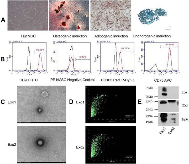
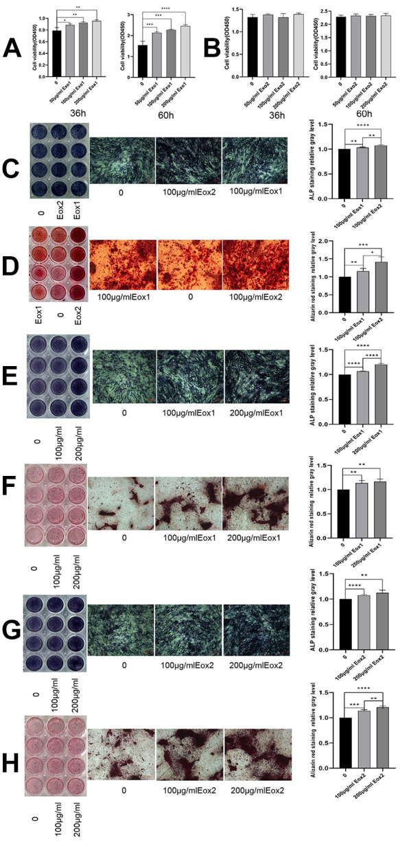
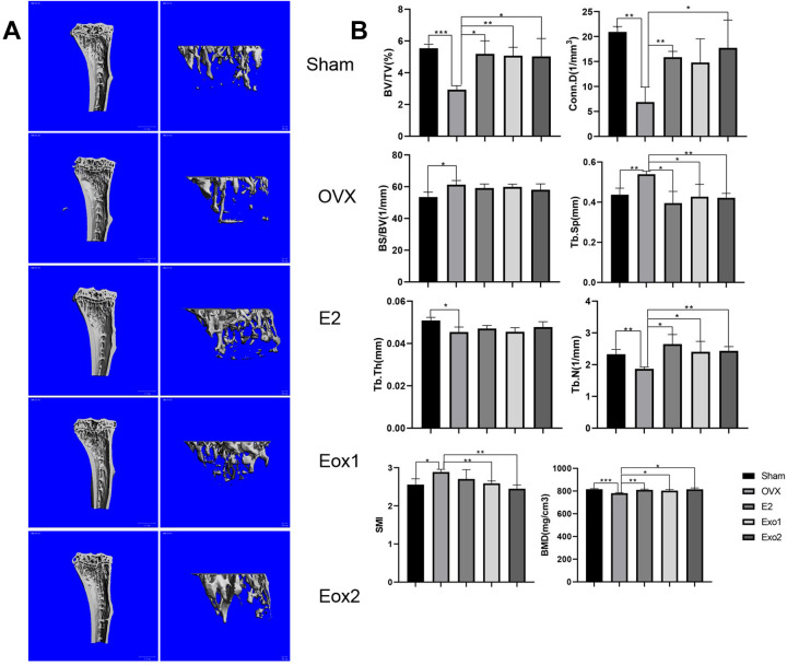
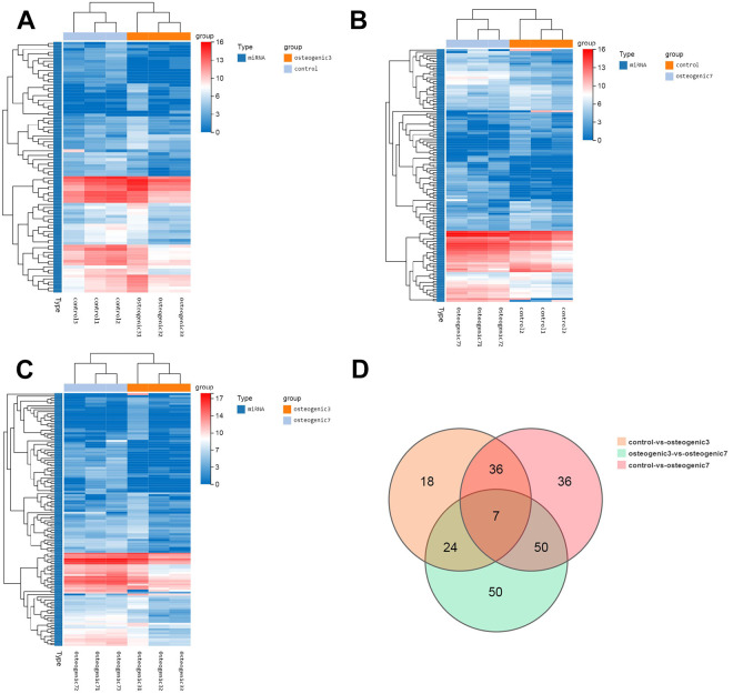
Mesenchymal stem cell (MSC) exosomes promote tissue regeneration and repair, and thus might be used to treat many diseases; however, the influence of microenvironmental conditions on exosomes remains unclear. The present study aimed to analyze the effect of osteogenic induction on the functions of human umbilical cord MSC (HucMSC)-derived exosomes. Exosomes from standardized stem cell culture (Exo1) and osteogenic differentiation-exosomes (Exo2) were co-cultured with osteoblasts, separately. Cell counting kit-8 assays, alkaline phosphatase and alizarin red staining were used to observe the exosomes' effects on osteoblast proliferation and differentiation. The levels of osteogenic differentiation-related proteins were analyzed using western blotting. Estrogen-deficient osteoporosis model mice were established, and treated with the two exosome preparations. Micro-computed tomography and hematoxylin and eosin staining were performed after 6 weeks. MicroRNAs in Exo1 and Exo2 were sequenced and analyzed using bioinformatic analyses. Compared with Exo1 group, Exo2 had a stronger osteogenic differentiation promoting effect, but a weaker proliferation promoting effect. In ovariectomy-induced osteoporosis mice, both Exo1 and Exo2 improved the tibial density and reversed osteoporosis in vivo. High-throughput microRNA sequencing identified 221 differentially expressed microRNAs in HucMSC-derived exosomes upon osteogenic induction as compared with the untreated control group. Importantly, we found that 41 of these microRNAs are potentially critical for MSC-secreted exosomes during osteogenic induction. Mechanistically, exosomal miRNAs derived from osteogenic induced-HucMSCs are involved in bone development and differentiation, such as osteoclast differentiation and the MAPK signaling pathway. The expression of hsa-mir-2110 and hsa-mir-328-3p gradually increased with prolonged osteogenic differentiation and regulated target genes associated with bone differentiation, suggesting that they are probably the most important osteogenesis regulatory microRNAs in exosomes. In conclusion, we examined the contribution of osteogenic induction to the function of exosomes secreted by HucMSCs following osteogenic differentiation in vitro and in vivo, and reveal the underlying molecular mechanisms of exosome action during osteoporosis.
间充质干细胞(MSC)外泌体促进组织再生和修复,因此可能用于治疗许多疾病;然而,微环境条件对外泌体的影响尚不清楚。本研究旨在分析成骨诱导对人脐带 MSC(HucMSC)衍生外泌体功能的影响。将来自标准化干细胞培养的外泌体(Exo1)和成骨分化外泌体(Exo2)分别与成骨细胞共培养。使用细胞计数试剂盒-8 测定、碱性磷酸酶和茜素红染色观察外泌体对成骨细胞增殖和分化的影响。使用 Western blot 分析成骨分化相关蛋白的水平。建立去势骨质疏松症模型小鼠,并分别用两种外泌体制剂处理。6 周后进行微计算机断层扫描和苏木精和伊红染色。使用生物信息学分析对 Exo1 和 Exo2 中的 microRNAs 进行测序和分析。与 Exo1 组相比,Exo2 具有更强的促骨分化作用,但促增殖作用较弱。在去势诱导的骨质疏松症小鼠中,Exo1 和 Exo2 均改善了胫骨密度并在体内逆转了骨质疏松症。高通量 microRNA 测序鉴定出与未处理对照组相比,成骨诱导后 HucMSC 衍生外泌体中有 221 个差异表达的 microRNAs。重要的是,我们发现这些 microRNAs 中有 41 个可能在 MSC 分泌的外泌体成骨诱导过程中是关键的。在机制上,成骨诱导的 HucMSC 衍生的外泌体中的 microRNAs 参与骨发育和分化,如破骨细胞分化和 MAPK 信号通路。hsa-mir-2110 和 hsa-mir-328-3p 的表达随着成骨分化时间的延长而逐渐增加,并调节与骨分化相关的靶基因,提示它们可能是外泌体中最重要的骨生成调节 microRNAs。总之,我们研究了成骨诱导对体外和体内 HucMSC 分泌的外泌体功能的影响,并揭示了外泌体在骨质疏松症过程中作用的潜在分子机制。