Department of Psychiatry and Psychology, Mayo Clinic Depression Center, Mayo Clinic, 200 1st St SW, Rochester, MN, 55905, United States.
Institute for Mental and Physical Health and Clinical Translation (IMPACT), Metabolic Research Unit, School of Medicine, Deakin University, 75 Pigdons Road, Waurn Ponds, VIC, 3216, Australia.
Transl Psychiatry. 2021 Nov 25;11(1):598. doi: 10.1038/s41398-021-01716-w.
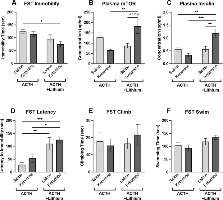
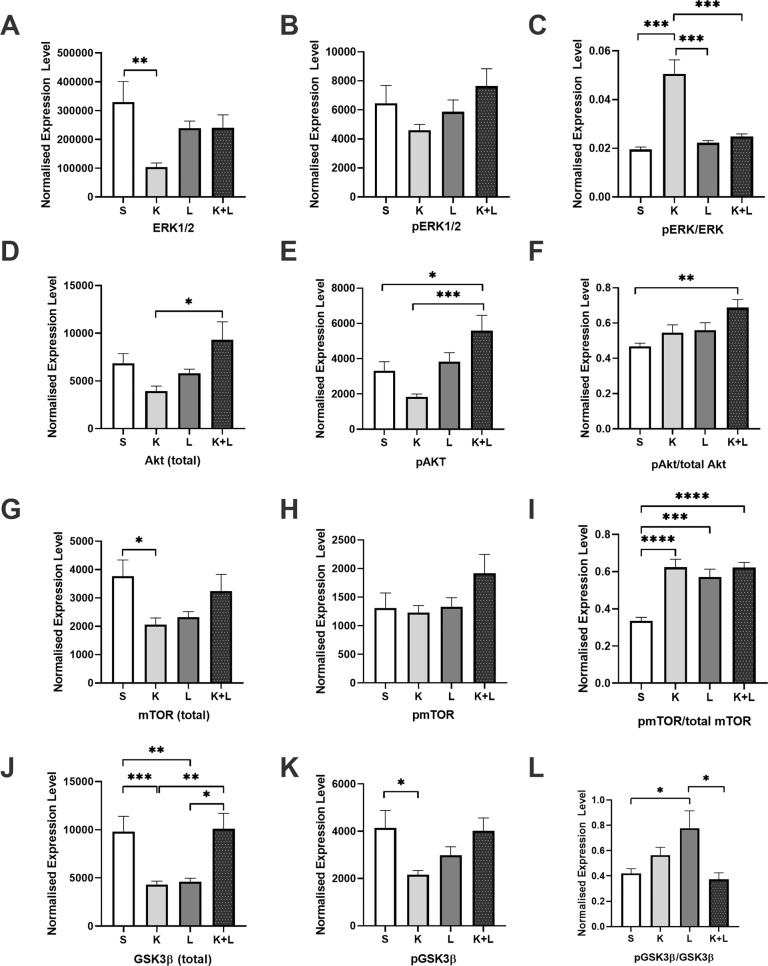
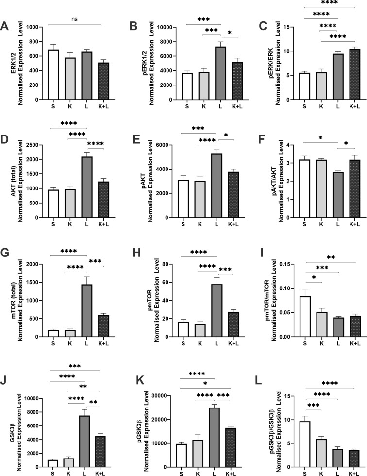
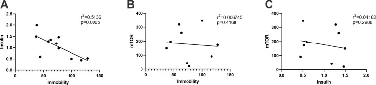
Lithium, a mood stabilizer and common adjunctive treatment for refractory depression, shares overlapping mechanisms of action with ketamine and enhances the duration of ketamine's antidepressant actions in rodent models at sub-therapeutic doses. Yet, in a recent clinical trial, lithium co-treatment with ketamine failed to improve antidepressant outcomes in subjects previously shown to respond to ketamine alone. The potential for lithium augmentation to improve antidepressant outcomes in ketamine nonresponders, however, has not been explored. The current study examined the behavioral, molecular and metabolic actions of lithium and ketamine co-treatment in a rodent model of antidepressant resistance. Male Wistar rats were administered adrenocorticotropic hormone (ACTH; 100 µg/day, i.p. over 14 days) and subsequently treated with ketamine (10 mg/kg; 2 days; n = 12), lithium (37 mg/kg; 2 days; n = 12), ketamine + lithium (10 mg/kg + 37 mg/kg; 2 days; n = 12), or vehicle saline (0.9%; n = 12). Rats were subjected to open field (6 min) and forced swim tests (6 min). Peripheral blood and brain prefrontal cortical (PFC) tissue was collected one hour following stress exposure. Western blotting was used to determine the effects of treatment on extracellular signal-regulated kinase (ERK); mammalian target of rapamycin (mTOR), phospho kinase B (Akt), and glycogen synthase kinase-3ß (GSK3ß) protein levels in the infralimbic (IL) and prelimbic (PL) subregions of the PFC. Prefrontal oxygen consumption rate (OCR) and extracellular acidification rates (ECAR) were also determined in anterior PFC tissue at rest and following stimulation with brain-derived neurotrophic factor (BDNF) and tumor necrosis factor α (TNFα). Blood plasma levels of mTOR and insulin were determined using enzyme-linked immunosorbent assays (ELISAs). Overall, rats receiving ketamine+lithium displayed a robust antidepressant response to the combined treatment as demonstrated through significant reductions in immobility time (p < 0.05) and latency to immobility (p < 0.01). These animals also had higher expression of plasma mTOR (p < 0.01) and insulin (p < 0.001). Tissue bioenergetics analyses revealed that combined ketamine+lithium treatment did not significantly alter the respiratory response to BDNF or TNFα. Animals receiving both ketamine and lithium had significantly higher phosphorylation (p)-to-total expression ratios of mTOR (p < 0.001) and Akt (p < 0.01), and lower ERK in the IL compared to control animals. In contrast, pmTOR/mTOR levels were reduced in the PL of ketamine+lithium treated animals, while pERK/ERK expression levels were elevated. Taken together, these data demonstrate that lithium augmentation of ketamine in antidepressant nonresponsive animals improves antidepressant-like behavioral responses under stress, together with peripheral insulin efflux and region-specific PFC insulin signaling.
锂,一种心境稳定剂,常用于难治性抑郁症的辅助治疗,与氯胺酮有重叠的作用机制,并在亚治疗剂量下增强氯胺酮在啮齿动物模型中的抗抑郁作用持续时间。然而,在最近的一项临床试验中,锂与氯胺酮联合治疗未能改善先前对氯胺酮有反应的患者的抗抑郁结果。然而,锂增强治疗在氯胺酮无反应者中改善抗抑郁结果的潜力尚未得到探索。本研究在抗抑郁药物抵抗的啮齿动物模型中研究了锂和氯胺酮联合治疗的行为、分子和代谢作用。雄性 Wistar 大鼠给予促肾上腺皮质激素(ACTH;100μg/天,腹腔内注射 14 天),随后用氯胺酮(10mg/kg;2 天;n=12)、锂(37mg/kg;2 天;n=12)、氯胺酮+锂(10mg/kg+37mg/kg;2 天;n=12)或生理盐水(0.9%;n=12)治疗。大鼠进行了旷场(6 分钟)和强迫游泳试验(6 分钟)。应激暴露后 1 小时采集外周血和大脑前额皮质(PFC)组织。Western blot 用于确定治疗对 PFC 下额(IL)和前额(PL)亚区细胞外信号调节激酶(ERK);哺乳动物雷帕霉素靶蛋白(mTOR)、磷酸化蛋白激酶 B(Akt)和糖原合酶激酶-3β(GSK3β)蛋白水平的影响。在前额皮质组织中还测定了静息时和脑源性神经营养因子(BDNF)和肿瘤坏死因子α(TNFα)刺激后的氧消耗率(OCR)和细胞外酸化率(ECAR)。使用酶联免疫吸附测定(ELISA)测定血浆 mTOR 和胰岛素水平。总的来说,接受氯胺酮+锂治疗的大鼠对联合治疗表现出强烈的抗抑郁反应,表现为不动时间显著减少(p<0.05)和不动潜伏期显著降低(p<0.01)。这些动物的血浆 mTOR(p<0.01)和胰岛素(p<0.001)水平也更高。组织生物能量学分析表明,氯胺酮+锂联合治疗并未显著改变 BDNF 或 TNFα 的呼吸反应。与对照动物相比,接受氯胺酮和锂的动物在 IL 中 mTOR(p<0.001)和 Akt(p<0.01)的磷酸化(p)与总表达比显著升高,ERK 降低。相比之下,氯胺酮+锂治疗动物的 PL 中 pmTOR/mTOR 水平降低,而 pERK/ERK 表达水平升高。总之,这些数据表明,锂增强抗抑郁药物抵抗动物的氯胺酮治疗可改善应激下的抗抑郁样行为反应,以及外周胰岛素流出和特定区域的 PFC 胰岛素信号。