Borm Kai J, Kleine Vennekate Johanne, Vagedes Jan, Islam Mohammad O A, Duma Marciana N, Loos Maximilian, Combs Stephanie E, Schiller Kilian, Klusen Sophie, Paepke Stefan, Kiechle Marion B, Paepke Daniela
Department of Radiation Oncology, Klinikum Rechts der Isar, Medical School, Technical University Munich, 81675 Munich, Germany.
ARCIM Institute (Academic Research in Complementary and Integrative Medicine), 70794 Filderstadt, Germany.
Cancers (Basel). 2021 Nov 20;13(22):5826. doi: 10.3390/cancers13225826.
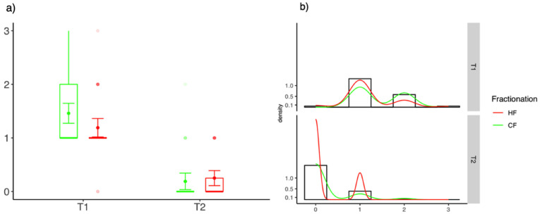
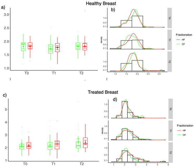
The current study aims to determine whether hypofractionated radiotherapy (HF) leads to lower rates of acute radiodermatitis compared to conventional normofractionated radiotherapy (CF). A total of 166 patients with invasive breast cancer or DCIS were included in a prospective cohort study. Evaluation of acute radiodermatitis was obtained before radiotherapy, at the end of the treatment (T1), and 6 weeks after the treatment (T2) using CTCAE (v5.0) scores, the Skindex-16 questionnaire, and ultrasound measurement of the skin. CTCAE and Skindex-16 scores in the CF-group were significantly higher compared to the HF group indicating more pronounced side effects at the end of the treatment (CTCAE: CF-RT 1.0 (IQR: 0.0) vs. HF-RT 0.0 (0.25); = 0.03; Skindex-16: CF: 20.8 (IQR: 25.8); HF: 8.3 (27.1); = 0.04). At 6 weeks after the treatment, no significant differences between the two fractionation schemes were observed. Ultrasound based assessment showed that the skin thickness in the treated breast was higher compared to the healthy breast at all time-points. However, no significant difference between HF and CF was seen either at T1 or T2. The current study complements and confirms pre-existing evidence that HF leads to a lower degree of acute radiodermatitis and better patient reported outcome compared to CF at the end of treatment. This should be considered whenever fractionation of adjuvant breast cancer treatment is being discussed.
本研究旨在确定与传统的常规分割放疗(CF)相比,大分割放疗(HF)是否会导致更低的急性放射性皮炎发生率。一项前瞻性队列研究纳入了166例浸润性乳腺癌或导管原位癌患者。在放疗前、治疗结束时(T1)以及治疗后6周(T2),使用CTCAE(v5.0)评分、Skindex-16问卷以及皮肤超声测量对急性放射性皮炎进行评估。CF组的CTCAE和Skindex-16评分显著高于HF组,表明在治疗结束时副作用更明显(CTCAE:CF-RT 1.0(IQR:0.0) vs. HF-RT 0.0(0.25);P = 0.03;Skindex-16:CF:20.8(IQR:25.8);HF:8.3(27.1);P = 0.04)。在治疗后6周,未观察到两种分割方案之间存在显著差异。基于超声的评估显示,在所有时间点,治疗侧乳房的皮肤厚度均高于健康侧乳房。然而,在T1或T2时,HF和CF之间也未观察到显著差异。本研究补充并证实了先前已有的证据,即与CF相比,HF在治疗结束时导致的急性放射性皮炎程度更低,患者报告的结局更好。在讨论辅助性乳腺癌治疗的分割方式时,应考虑这一点。