Granata Vincenza, Ianniello Stefania, Fusco Roberta, Urraro Fabrizio, Pupo Davide, Magliocchetti Simona, Albarello Fabrizio, Campioni Paolo, Cristofaro Massimo, Di Stefano Federica, Fusco Nicoletta, Petrone Ada, Schininà Vincenzo, Villanacci Alberta, Grassi Francesca, Grassi Roberta, Grassi Roberto
Division of Radiology, Istituto Nazionale Tumori IRCCS Fondazione Pascale-IRCCS di Napoli, 80131 Naples, Italy.
Radiology Unit, National Institute for Infectious Diseases Lazzaro Spallanzani IRCCS, 00149 Rome, Italy.
J Pers Med. 2021 Oct 28;11(11):1103. doi: 10.3390/jpm11111103.
To investigate two commercial software and their efficacy in the assessment of chest CT sequelae in patients affected by COVID-19 pneumonia, comparing the consistency of tools.
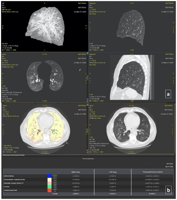
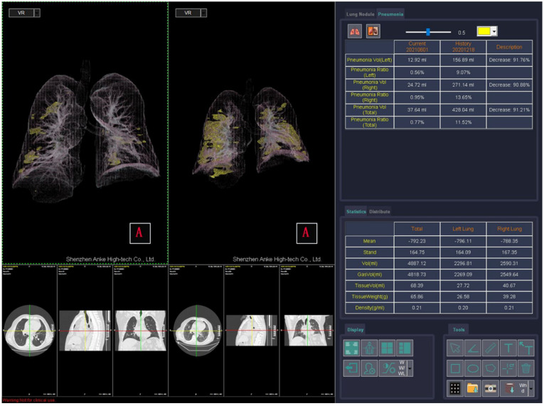
Included in the study group were 120 COVID-19 patients (56 women and 104 men; 61 years of median age; range: 21-93 years) who underwent chest CT examinations at discharge between 5 March 2020 and 15 March 2021 and again at a follow-up time (3 months; range 30-237 days). A qualitative assessment by expert radiologists in the infectious disease field (experience of at least 5 years) was performed, and a quantitative evaluation using thoracic VCAR software (GE Healthcare, Chicago, Illinois, United States) and a pneumonia module of ANKE ASG-340 CT workstation (HTS Med & Anke, Naples, Italy) was performed. The qualitative evaluation included the presence of ground glass opacities (GGOs) consolidation, interlobular septal thickening, fibrotic-like changes (reticular pattern and/or honeycombing), bronchiectasis, air bronchogram, bronchial wall thickening, pulmonary nodules surrounded by GGOs, pleural and pericardial effusion, lymphadenopathy, and emphysema. A quantitative evaluation included the measurements of GGOs, consolidations, emphysema, residual healthy parenchyma, and total lung volumes for the right and left lung. A chi-square test and non-parametric test were utilized to verify the differences between groups. Correlation coefficients were used to analyze the correlation and variability among quantitative measurements by different computer tools. A receiver operating characteristic (ROC) analysis was performed.
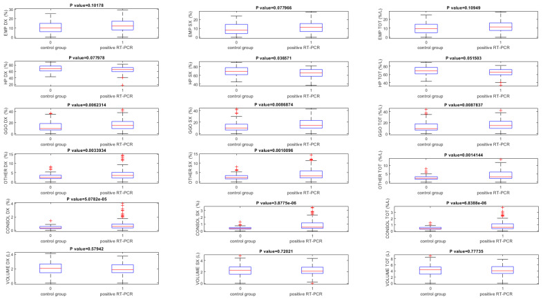
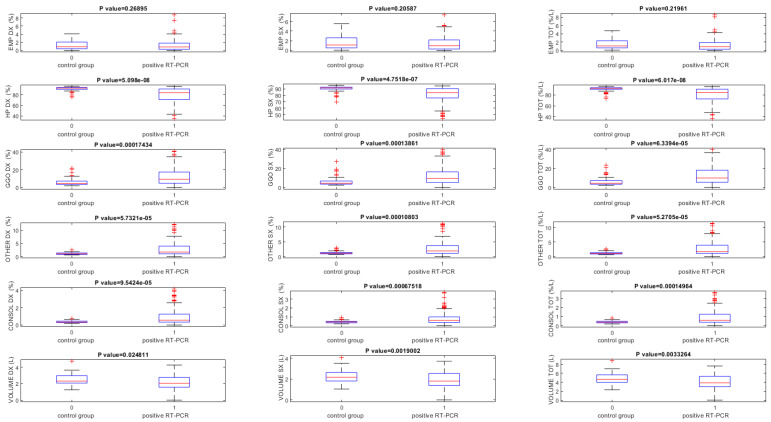
The correlation coefficients showed great variability among the quantitative measurements by different tools when calculated on baseline CT scans and considering all patients. Instead, a good correlation (≥0.6) was obtained for the quantitative GGO, as well as the consolidation volumes obtained by two tools when calculated on baseline CT scans, considering the control group. An excellent correlation (≥0.75) was obtained for the quantitative residual healthy lung parenchyma volume, GGO, consolidation volumes obtained by two tools when calculated on follow-up CT scans, and for residual healthy lung parenchyma and GGO quantification when the percentage change of these volumes were calculated between a baseline and follow-up scan. The highest value of accuracy to identify patients with RT-PCR positive compared to the control group was obtained by a GGO total volume quantification by thoracic VCAR (accuracy = 0.75).
Computer aided quantification could be an easy and feasible way to assess chest CT sequelae due to COVID-19 pneumonia; however, a great variability among measurements provided by different tools should be considered.
研究两款商业软件及其在评估新冠病毒肺炎患者胸部CT后遗症方面的效果,比较工具之间的一致性。
研究组纳入了120例新冠病毒肺炎患者(56例女性和104例男性;中位年龄61岁;范围:21 - 93岁),这些患者于2020年3月5日至2021年3月15日出院时接受了胸部CT检查,并在随访期(3个月;范围30 - 237天)再次接受检查。由传染病领域至少有5年经验的放射科专家进行定性评估,并使用胸部VCAR软件(美国伊利诺伊州芝加哥通用电气医疗集团)和ANKE ASG - 340 CT工作站(意大利那不勒斯HTS Med & Anke)的肺炎模块进行定量评估。定性评估包括磨玻璃影(GGO)实变、小叶间隔增厚、纤维化样改变(网状影和/或蜂窝状影)、支气管扩张、空气支气管征、支气管壁增厚、GGO包绕的肺结节、胸腔和心包积液、淋巴结肿大以及肺气肿。定量评估包括测量左右肺的GGO、实变、肺气肿、残余健康实质和总肺容积。采用卡方检验和非参数检验来验证组间差异。相关系数用于分析不同计算机工具定量测量之间的相关性和变异性。进行了受试者操作特征(ROC)分析。
在基线CT扫描并考虑所有患者时,不同工具的定量测量之间相关系数显示出很大变异性。相反,在考虑对照组时,对于定量GGO以及两款工具在基线CT扫描时获得的实变容积,获得了良好的相关性(≥0.6)。对于定量残余健康肺实质容积、GGO以及两款工具在随访CT扫描时获得的实变容积,以及在计算这些容积在基线和随访扫描之间的百分比变化时的残余健康肺实质和GGO定量,获得了极好的相关性(≥0.75)。通过胸部VCAR对GGO总体积进行定量,与对照组相比,识别RT - PCR阳性患者的准确率最高(准确率 = 0.75)。
计算机辅助定量可能是评估新冠病毒肺炎胸部CT后遗症的一种简便可行的方法;然而,应考虑不同工具提供的测量结果之间存在很大变异性。