Department of Pharmacology, School of Pharmaceutical, Guangzhou University of Chinese Medicine, 232, Waihuan East Road, Guangzhou Higher Education Mega Center, Panyu District, Guangzhou, 510000, China.
Department of Stomatology, The First Affiliated Hospital, The School of Dental Medicine, Jinan University, Guangzhou, China.
J Nanobiotechnology. 2021 Nov 27;19(1):396. doi: 10.1186/s12951-021-01137-3.
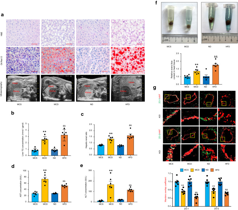
A recent study has reported that patients with nonalcoholic fatty liver disease (NAFLD) are more susceptible to coronary microvascular dysfunction (CMD), which may predict major adverse cardiac events. However, little is known regarding the causes of CMD during NAFLD. In this study, we aimed to explore the role of hepatic small extracellular vesicles (sEVs) in regulating the endothelial dysfunction of coronary microvessels during NAFLD.
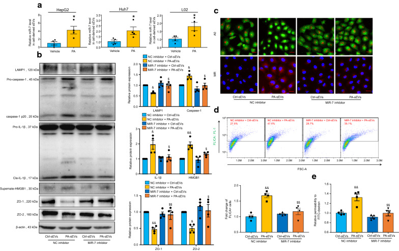
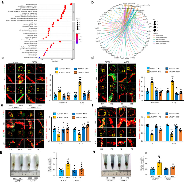
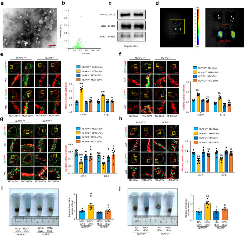
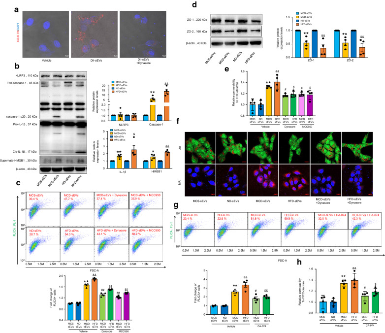
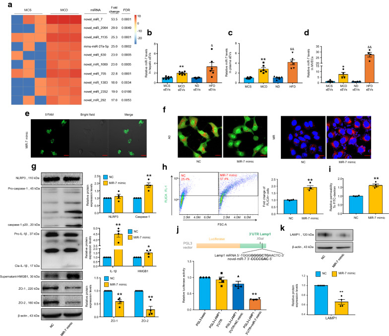
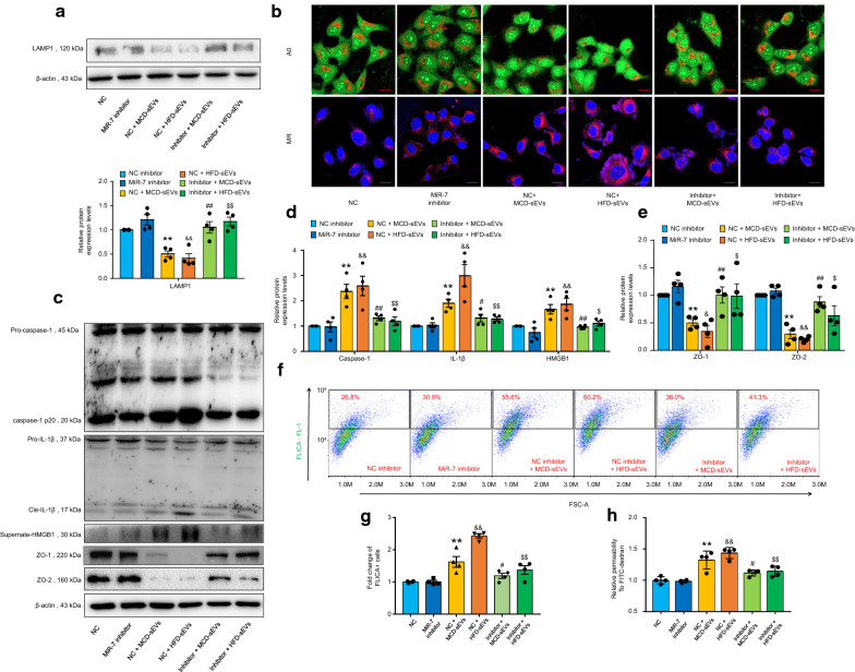
We established two murine NAFLD models by feeding mice a methionine-choline-deficient (MCD) diet for 4 weeks or a high-fat diet (HFD) for 16 weeks. We found that the NOD-like receptor family, pyrin domain containing 3 (NLRP3) inflammasome-dependent endothelial hyperpermeability occurred in coronary microvessels during both MCD diet and HFD-induced NAFLD. The in vivo and in vitro experiments proved that novel-microRNA(miR)-7-abundant hepatic sEVs were responsible for NLRP3 inflammasome-dependent endothelial barrier dysfunction. Mechanistically, novel-miR-7 directly targeted lysosomal associated membrane protein 1 (LAMP1) and promotes lysosomal membrane permeability (LMP), which in turn induced Cathepsin B-dependent NLRP3 inflammasome activation and microvascular endothelial hyperpermeability. Conversely, a specific novel-miR-7 inhibitor markedly improved endothelial barrier integrity. Finally, we proved that steatotic hepatocyte was a significant source of novel-miR-7-contained hepatic sEVs, and steatotic hepatocyte-derived sEVs were able to promote NLRP3 inflammasome-dependent microvascular endothelial hyperpermeability through novel-miR-7.
Hepatic sEVs contribute to endothelial hyperpermeability in coronary microvessels by delivering novel-miR-7 and targeting the LAMP1/Cathepsin B/NLRP3 inflammasome axis during NAFLD. Our study brings new insights into the liver-to-microvessel cross-talk and may provide a new diagnostic biomarker and treatment target for microvascular complications of NAFLD.
最近的一项研究表明,非酒精性脂肪性肝病(NAFLD)患者更容易发生冠状动脉微血管功能障碍(CMD),这可能预示着主要不良心脏事件的发生。然而,关于 NAFLD 期间 CMD 的原因知之甚少。在这项研究中,我们旨在探讨肝小细胞外囊泡(sEVs)在调节 NAFLD 期间冠状动脉微血管内皮功能障碍中的作用。
我们通过用蛋氨酸-胆碱缺乏(MCD)饮食喂养小鼠 4 周或高脂肪饮食(HFD)喂养 16 周建立了两种小鼠 NAFLD 模型。我们发现,NOD 样受体家族,富含吡喃结构域的 3(NLRP3)炎性小体依赖性内皮通透性增加发生在 MCD 饮食和 HFD 诱导的 NAFLD 期间的冠状动脉微血管中。体内和体外实验证明,新型 microRNA(miR)-7 丰富的肝 sEVs 是 NLRP3 炎性小体依赖性内皮屏障功能障碍的原因。从机制上讲,新型-miR-7 直接靶向溶酶体相关膜蛋白 1(LAMP1)并促进溶酶体膜通透性(LMP),进而诱导组织蛋白酶 B 依赖性 NLRP3 炎性小体激活和微血管内皮通透性增加。相反,特异性新型-miR-7 抑制剂可显著改善内皮屏障完整性。最后,我们证明脂肪变性肝细胞是新型-miR-7 含量丰富的肝 sEV 的重要来源,脂肪变性肝细胞衍生的 sEV 能够通过新型-miR-7 促进 NLRP3 炎性小体依赖性微血管内皮通透性增加。
在 NAFLD 期间,肝 sEV 通过递送新型-miR-7 并靶向 LAMP1/Cathepsin B/NLRP3 炎性小体轴,导致冠状动脉微血管内皮通透性增加。我们的研究为肝脏与微血管的相互作用提供了新的见解,并可能为 NAFLD 的微血管并发症提供新的诊断生物标志物和治疗靶点。