• 文献检索
  • 文档翻译
  • 深度研究
  • 学术资讯
  • Suppr Zotero 插件Zotero 插件
  • 邀请有礼
  • 套餐&价格
  • 历史记录
应用&插件
Suppr Zotero 插件Zotero 插件浏览器插件Mac 客户端Windows 客户端微信小程序
定价
高级版会员购买积分包购买API积分包
服务
文献检索文档翻译深度研究API 文档MCP 服务
关于我们
关于 Suppr公司介绍联系我们用户协议隐私条款
关注我们

Suppr 超能文献

核心技术专利:CN118964589B侵权必究
粤ICP备2023148730 号-1Suppr @ 2026

文献检索

告别复杂PubMed语法,用中文像聊天一样搜索,搜遍4000万医学文献。AI智能推荐,让科研检索更轻松。

立即免费搜索

文件翻译

保留排版,准确专业,支持PDF/Word/PPT等文件格式,支持 12+语言互译。

免费翻译文档

深度研究

AI帮你快速写综述,25分钟生成高质量综述,智能提取关键信息,辅助科研写作。

立即免费体验

杜氏利什曼原虫驱动的皮肤利什曼病创面中独特的微生物组谱和生物膜。

Distinct microbiome profiles and biofilms in Leishmania donovani-driven cutaneous leishmaniasis wounds.

机构信息

Department of Parasitology, Faculty of Medical Sciences, University of Sri Jayewardenepura, Gangodawila, Nugegoda, Sri Lanka.

Division of Pharmacy and Optometry, School of Health Sciences, Faculty of Biology, Medicine and Health, The University of Manchester, Manchester, UK.

出版信息

Sci Rep. 2021 Nov 30;11(1):23181. doi: 10.1038/s41598-021-02388-8.

DOI:10.1038/s41598-021-02388-8
PMID:34848752
原文链接:https://pmc.ncbi.nlm.nih.gov/articles/PMC8633208/
Abstract

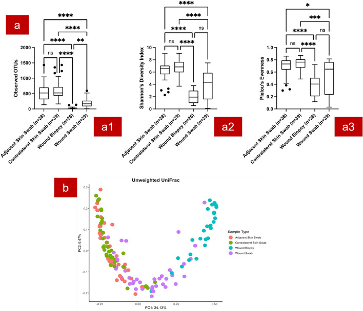
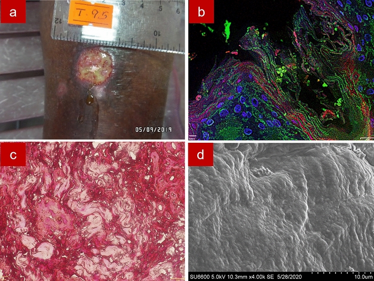
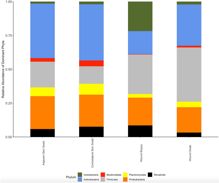
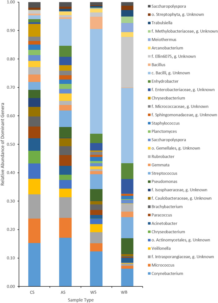
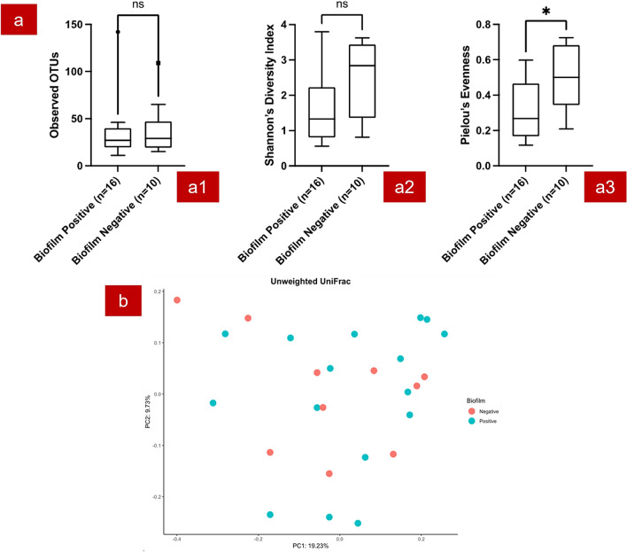
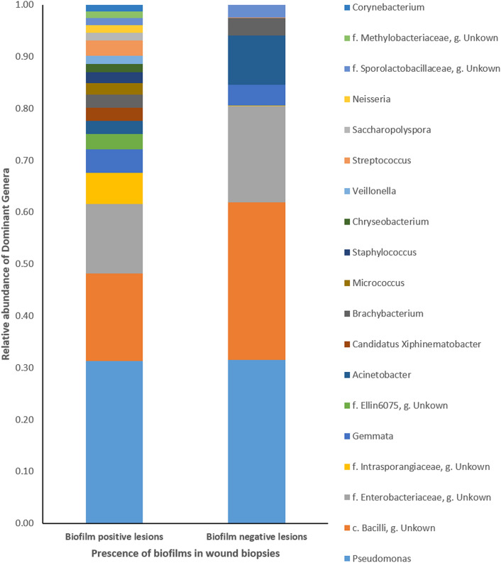
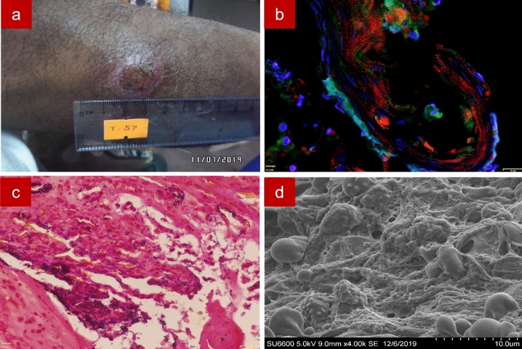
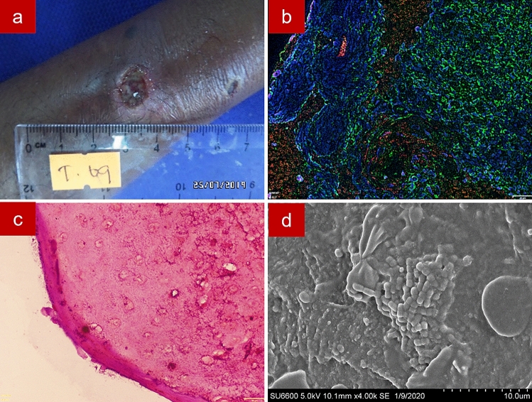
The endemic strain of Leishmania donovani in Sri Lanka causes cutaneous leishmaniasis (CL) rather than more common visceral form. We have visualized biofilms and profiled the microbiome of lesions and unaffected skin in thirty-nine CL patients. Twenty-four lesions (61.5%) were biofilm-positive according to fluorescence in situ hybridization. Biopsies of biofilm-positive lesions were dominated by Pseudomonas, class Bacilli and Enterobacteriaceae and distinguished by significantly lower community evenness. Higher relative abundance of a class Bacilli OTU was detected in wound swabs versus contralateral skin. Wound swabs and biopsies had significantly distinct microbiome profiles and lower diversity compared to unaffected skin. Greater abundances of potentially pathogenic organisms were observed in wet ulcers, lesions with high parasite loads and large wounds. In summary, more than half of L. donovani associated CL wounds harboured biofilms and the wounds exhibited a distinct, less diverse, microbiome than unaffected skin.

摘要

斯里兰卡地方性的杜氏利什曼原虫(Leishmania donovani)株引起皮肤利什曼病(cutaneous leishmaniasis,CL),而不是更为常见的内脏利什曼病。我们对 39 例 CL 患者的病变和未受影响的皮肤进行了生物膜可视化和微生物组分析。根据荧光原位杂交,有 24 处病变(61.5%)呈生物膜阳性。生物膜阳性病变的活检标本主要由假单胞菌、B 类杆菌和肠杆菌科组成,其群落均匀度显著降低。与对侧皮肤相比,在伤口拭子中检测到 B 类杆菌 OTU 的相对丰度更高。与未受影响的皮肤相比,伤口拭子和活检标本的微生物组谱明显不同,多样性也较低。在湿性溃疡、寄生虫负荷高和大伤口的病变中,观察到更多潜在的致病性生物体。总之,超过一半的与杜氏利什曼原虫相关的 CL 伤口存在生物膜,并且与未受影响的皮肤相比,伤口的微生物组具有明显不同、多样性较低的特征。

https://cdn.ncbi.nlm.nih.gov/pmc/blobs/411e/8633208/13bc2360cc16/41598_2021_2388_Fig8_HTML.jpg
https://cdn.ncbi.nlm.nih.gov/pmc/blobs/411e/8633208/6e8bfbc89103/41598_2021_2388_Fig1_HTML.jpg
https://cdn.ncbi.nlm.nih.gov/pmc/blobs/411e/8633208/524c79cdb89b/41598_2021_2388_Fig2_HTML.jpg
https://cdn.ncbi.nlm.nih.gov/pmc/blobs/411e/8633208/f169bfb71a2f/41598_2021_2388_Fig3_HTML.jpg
https://cdn.ncbi.nlm.nih.gov/pmc/blobs/411e/8633208/a9fc61181c29/41598_2021_2388_Fig4_HTML.jpg
https://cdn.ncbi.nlm.nih.gov/pmc/blobs/411e/8633208/0cffa259c96a/41598_2021_2388_Fig5_HTML.jpg
https://cdn.ncbi.nlm.nih.gov/pmc/blobs/411e/8633208/cd51198c58b2/41598_2021_2388_Fig6_HTML.jpg
https://cdn.ncbi.nlm.nih.gov/pmc/blobs/411e/8633208/4423b06fbaae/41598_2021_2388_Fig7_HTML.jpg
https://cdn.ncbi.nlm.nih.gov/pmc/blobs/411e/8633208/13bc2360cc16/41598_2021_2388_Fig8_HTML.jpg

相似文献

1
Distinct microbiome profiles and biofilms in Leishmania donovani-driven cutaneous leishmaniasis wounds.杜氏利什曼原虫驱动的皮肤利什曼病创面中独特的微生物组谱和生物膜。
Sci Rep. 2021 Nov 30;11(1):23181. doi: 10.1038/s41598-021-02388-8.
2
Genetic diversity of Leishmania donovani that causes cutaneous leishmaniasis in Sri Lanka: a cross sectional study with regional comparisons.斯里兰卡引起皮肤利什曼病的杜氏利什曼原虫的遗传多样性:一项区域比较的横断面研究
BMC Infect Dis. 2017 Dec 22;17(1):791. doi: 10.1186/s12879-017-2883-x.
3
Diagnosing Cutaneous leishmaniasis using Fluorescence Hybridization: the Sri Lankan Perspective.应用荧光杂交技术诊断皮肤利什曼病:斯里兰卡视角。
Pathog Glob Health. 2019 Jun;113(4):180-190. doi: 10.1080/20477724.2019.1650228. Epub 2019 Aug 20.
4
Efficacy of a new rapid diagnostic test kit to diagnose Sri Lankan cutaneous leishmaniasis caused by Leishmania donovani.一种新型快速诊断试剂盒用于诊断由杜氏利什曼原虫引起的斯里兰卡皮肤利什曼病的疗效。
PLoS One. 2017 Nov 14;12(11):e0187024. doi: 10.1371/journal.pone.0187024. eCollection 2017.
5
Clinical and epidemiological studies on the cutaneous leishmaniasis caused by Leishmania (Leishmania) donovani in Sri Lanka.斯里兰卡杜氏利什曼原虫(利什曼原虫属)引起的皮肤利什曼病的临床和流行病学研究
Ann Trop Med Parasitol. 2010 Apr;104(3):213-23. doi: 10.1179/136485910X12647085215615.
6
First Serological Study Revealing High Humoral Response and Evidence for Antigenic Heterogeneity in Induced CL in Sri Lanka.首次血清学研究揭示了斯里兰卡诱导性 CL 中的高体液反应和抗原异质性证据。
Biomed Res Int. 2020 Sep 29;2020:5271657. doi: 10.1155/2020/5271657. eCollection 2020.
7
Polymerase chain reaction detection of Leishmania DNA in skin biopsy samples in Sri Lanka where the causative agent of cutaneous leishmaniasis is Leishmania donovani.在斯里兰卡,皮肤利什曼病的病原体为杜氏利什曼原虫,通过聚合酶链反应检测皮肤活检样本中的利什曼原虫DNA。
Mem Inst Oswaldo Cruz. 2015 Dec;110(8):1017-23. doi: 10.1590/0074-02760150286. Epub 2015 Dec 11.
8
Histopathology of Cutaneous Leishmaniasis Caused by in Sri Lanka.斯里兰卡引起的皮肤利什曼病的组织病理学。
Biomed Res Int. 2020 May 2;2020:4926819. doi: 10.1155/2020/4926819. eCollection 2020.
9
Clinical and epidemiological characteristics of cutaneous leishmaniasis in Sri Lanka.斯里兰卡皮肤利什曼病的临床和流行病学特征。
BMC Infect Dis. 2018 Mar 6;18(1):108. doi: 10.1186/s12879-018-2999-7.
10
First Evidence from Sri Lanka for Subphenotypic Diversity within -Induced Classical Cutaneous Leishmaniasis.斯里兰卡诱导的经典皮肤利什曼病亚表型多样性的首个证据。
Biomed Res Int. 2021 Jan 27;2021:3537968. doi: 10.1155/2021/3537968. eCollection 2021.

引用本文的文献

1
Biofilm Resilience: Molecular Mechanisms Driving Antibiotic Resistance in Clinical Contexts.生物膜弹性:临床环境中驱动抗生素耐药性的分子机制。
Biology (Basel). 2025 Feb 6;14(2):165. doi: 10.3390/biology14020165.
2
Parasite-microbiota interactions: a pathway to innovative interventions for Chagas disease, leishmaniasis, and ascariasis.寄生虫-微生物群相互作用:治疗恰加斯病、利什曼病和蛔虫病的创新干预途径。
Future Microbiol. 2025 Feb;20(2):149-161. doi: 10.1080/17460913.2024.2431417. Epub 2024 Nov 22.
3
Prokaryotic and eukaryotic skin microbiota modifications triggered by Leishmania infection in localized Cutaneous Leishmaniasis.

本文引用的文献

1
Biofilm prevalence and microbial characterisation in chronic wounds in a Sri Lankan cohort.斯里兰卡队列慢性伤口中生物膜的流行情况和微生物特征。
Lett Appl Microbiol. 2021 Oct;73(4):477-485. doi: 10.1111/lam.13532. Epub 2021 Jul 20.
2
The microbiome of diabetic foot ulcers: a comparison of swab and tissue biopsy wound sampling techniques using 16S rRNA gene sequencing.糖尿病足溃疡的微生物组:使用 16S rRNA 基因测序比较拭子和组织活检伤口采样技术。
BMC Microbiol. 2020 Jun 16;20(1):163. doi: 10.1186/s12866-020-01843-2.
3
Microbial predictors of healing and short-term effect of debridement on the microbiome of chronic wounds.
利什曼原虫感染引起局限性皮肤利什曼病时,原核生物和真核生物皮肤微生物组的改变。
PLoS Negl Trop Dis. 2024 Mar 13;18(3):e0012029. doi: 10.1371/journal.pntd.0012029. eCollection 2024 Mar.
4
Designing of a new multi-epitope vaccine against Leishmania major using Leish-F1 epitopes: An In-silico study.利用 Leish-F1 表位设计新型多表位疫苗对抗利什曼原虫:一项计算机研究。
PLoS One. 2024 Jan 2;19(1):e0295495. doi: 10.1371/journal.pone.0295495. eCollection 2024.
5
Unraveling the interplay between unicellular parasites and bacterial biofilms: Implications for disease persistence and antibiotic resistance.解析单细胞寄生虫与细菌生物膜的相互作用:对疾病持续存在和抗生素耐药性的影响。
Virulence. 2024 Dec;15(1):2289775. doi: 10.1080/21505594.2023.2289775. Epub 2023 Dec 6.
6
Multiomic profiling of cutaneous leishmaniasis infections reveals microbiota-driven mechanisms underlying disease severity.皮肤利什曼病感染的多组学分析揭示了疾病严重程度的微生物群驱动机制。
Sci Transl Med. 2023 Oct 18;15(718):eadh1469. doi: 10.1126/scitranslmed.adh1469.
7
Effect of Clindamycin on Intestinal Microbiome and Miltefosine Pharmacology in Hamsters Infected with .克林霉素对感染……的仓鼠肠道微生物群和米替福新药理学的影响
Antibiotics (Basel). 2023 Feb 9;12(2):362. doi: 10.3390/antibiotics12020362.
8
Possible clinical implications and future directions of managing bacterial biofilms in cutaneous leishmaniasis wounds.皮肤利什曼病伤口中细菌生物膜管理的潜在临床意义及未来方向。
Trop Med Health. 2022 Aug 26;50(1):58. doi: 10.1186/s41182-022-00455-y.
慢性伤口微生物组学清创术对愈合的微生物预测因子及短期影响
NPJ Biofilms Microbiomes. 2020 May 1;6(1):21. doi: 10.1038/s41522-020-0130-5.
4
Bacterial Diversity of Diabetic Foot Ulcers: Current Status and Future Prospectives.糖尿病足溃疡的细菌多样性:现状与未来展望
J Clin Med. 2019 Nov 10;8(11):1935. doi: 10.3390/jcm8111935.
5
Diagnosing Cutaneous leishmaniasis using Fluorescence Hybridization: the Sri Lankan Perspective.应用荧光杂交技术诊断皮肤利什曼病:斯里兰卡视角。
Pathog Glob Health. 2019 Jun;113(4):180-190. doi: 10.1080/20477724.2019.1650228. Epub 2019 Aug 20.
6
Coinfection with Leishmania major and Staphylococcus aureus enhances the pathologic responses to both microbes through a pathway involving IL-17A.感染利什曼原虫和金黄色葡萄球菌会通过涉及白细胞介素-17A 的途径增强对这两种微生物的病理反应。
PLoS Negl Trop Dis. 2019 May 20;13(5):e0007247. doi: 10.1371/journal.pntd.0007247. eCollection 2019 May.
7
Leishmaniasis.利什曼病。
Lancet. 2018 Sep 15;392(10151):951-970. doi: 10.1016/S0140-6736(18)31204-2. Epub 2018 Aug 17.
8
Bacterial Aggregates Establish at the Edges of Acute Epidermal Wounds.细菌聚集体在急性表皮伤口边缘形成。
Adv Wound Care (New Rochelle). 2018 Apr 1;7(4):105-113. doi: 10.1089/wound.2017.0770.
9
Measuring the microbiome of chronic wounds with use of a topical antimicrobial dressing - A feasibility study.使用局部抗菌敷料测量慢性伤口的微生物群——一项可行性研究。
PLoS One. 2017 Nov 20;12(11):e0187728. doi: 10.1371/journal.pone.0187728. eCollection 2017.
10
Consensus guidelines for the identification and treatment of biofilms in chronic nonhealing wounds.慢性难愈合伤口中生物膜识别与治疗的共识指南。
Wound Repair Regen. 2017 Sep;25(5):744-757. doi: 10.1111/wrr.12590. Epub 2017 Dec 12.