Reinstein Dan Z, Vida Ryan S, Archer Timothy J
London Vision Clinic, London, UK.
Columbia University Medical Center, New York, NY, USA.
Clin Ophthalmol. 2021 Nov 20;15:4485-4497. doi: 10.2147/OPTH.S330879. eCollection 2021.
Report the outcomes of the implantable collamer lens (ICL) in myopic astigmatism using very high-frequency (VHF) digital ultrasound sizing.
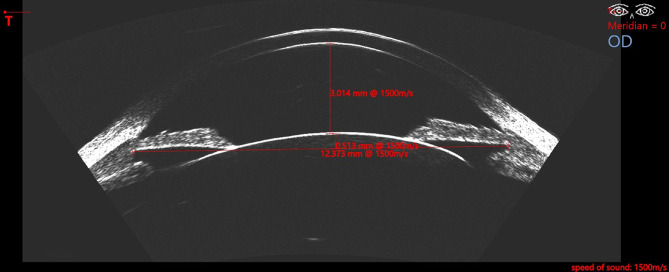
Analysis of 42 consecutive ICL procedures using EVO and EVO+ (Visian V4c) lenses (STAAR Surgical) was done. ICL size was chosen using the ultrasound-based Kojima Formula with Insight 100 VHF digital ultrasound (VHFDU). Standard visual outcomes analysis was performed using 3-month data, also including contrast sensitivity, refractive and corneal vector analysis, and ECC. Postoperative lens position was evaluated using VHF digital ultrasound.
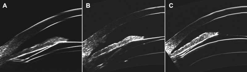
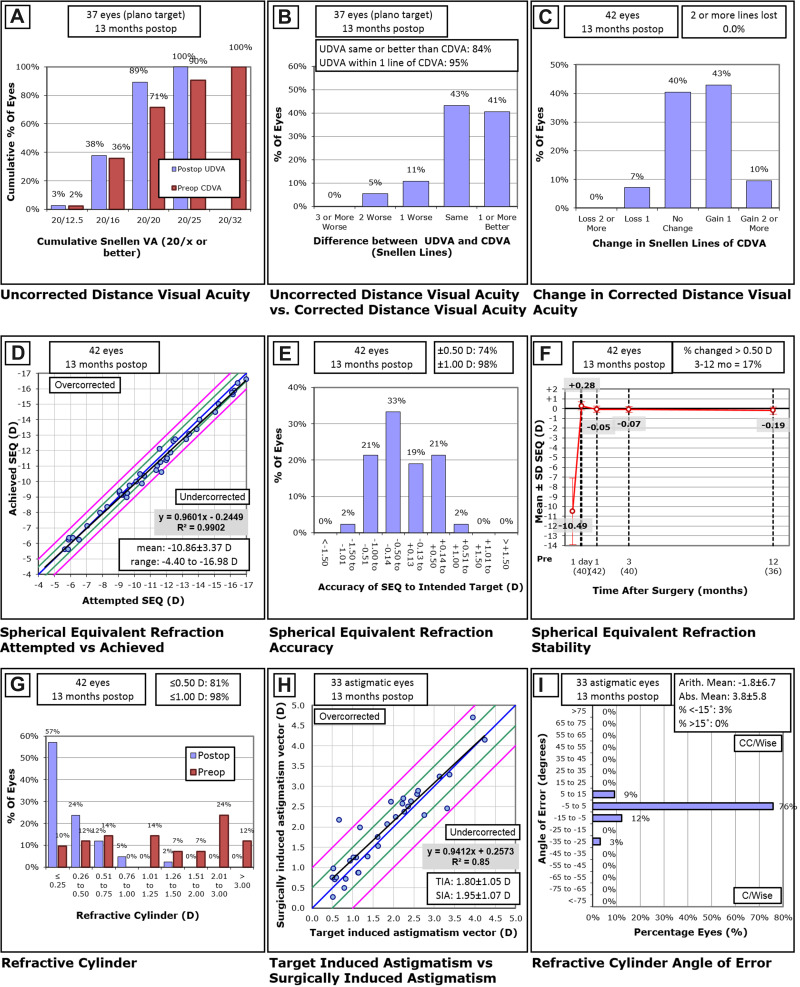
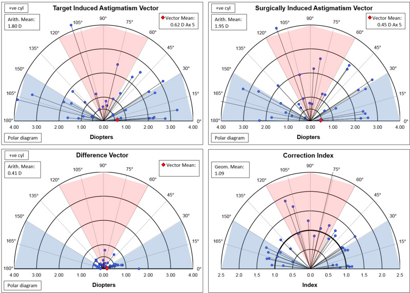
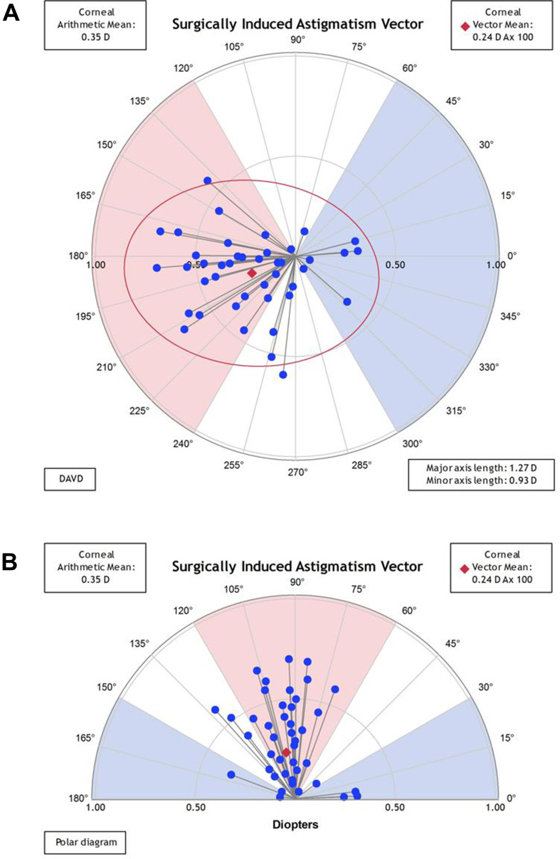
Attempted SEQ was -10.83±3.39D (-4.40 to -16.98D). Cylinder was -1.46±1.15D (0.00 to -4.25D). One-year follow-up was available in 86% of the eyes and 3 months in 96%. Postoperative UDVA was 20/20 or better in 89% of the eyes, relative to 71% preoperatively. Postoperative SEQ refraction was ±0.50 D in 74% and ±1.00 D in 98% of the eyes. There was a gain of one line of CDVA in 43% of the eyes, 2 or more lines in 10% of the eyes, while there was a one line loss in 7% and no eyes lost 2 or more lines. The vector mean for the corneal SIA was 0.24 D Ax 100. Contrast sensitivity showed a statistically significant increase with a mean of 0.14 log units at 6, 12, and 18 cycles per degree (P<0.01). The mean change in ECC was -153±353 cells/mm. Lens vault was 506±233 µm (114-924 µm). Footplate insertion was in zonular position in 48.3%, ciliary body in 49.2%, and sulcus in 2.5% of locations.
ICL implantation resulted in high safety and efficacy but with an implantation vault range that ideally would be improved upon. VHF digital ultrasound of the lens footplate and posterior anatomical relations may provide essential information for evaluating postoperative vault outliers.
报告使用超高频(VHF)数字超声测量法植入可植入式角膜接触镜(ICL)治疗近视散光的效果。
对连续42例使用EVO和EVO+(Visian V4c)镜片(STAAR Surgical公司)进行的ICL手术进行分析。使用基于超声的小岛公式和Insight 100 VHF数字超声(VHFDU)选择ICL尺寸。使用3个月的数据进行标准视力结果分析,还包括对比敏感度、屈光和角膜矢量分析以及角膜内皮细胞计数(ECC)。使用VHF数字超声评估术后晶状体位置。
预期等效球镜度(SEQ)为-10.83±3.39D(-4.40至-16.98D)。柱镜度为-1.46±1.15D(0.00至-4.25D)。86%的眼睛有1年随访数据,96%的眼睛有3个月随访数据。术后89%的眼睛最佳矫正视力(UDVA)达到20/20或更好,术前这一比例为71%。术后74%的眼睛SEQ屈光在±0.50 D以内,98%的眼睛在±1.00 D以内。43%的眼睛矫正视力(CDVA)提高了一行,10%的眼睛提高了两行或更多行,7%的眼睛下降了一行,没有眼睛下降两行或更多行。角膜手术性散光(SIA)的矢量平均值为0.24 D,轴位100。对比敏感度在每度6、12和18周期时平均提高0.14对数单位,差异有统计学意义(P<0.01)。ECC的平均变化为-153±353个细胞/mm。晶状体拱高为506±233 µm(114 - 924 µm)。48.3%的植入位置晶状体襻位于睫状小带,49.2%位于睫状体,2.5%位于睫状沟。
ICL植入具有高安全性和有效性,但植入拱高范围理想情况下仍有待改善。对晶状体襻和后部解剖关系进行VHF数字超声检查可能为评估术后拱高异常提供重要信息。