Knyazev Evgeny, Maltseva Diana, Raygorodskaya Maria, Shkurnikov Maxim
Laboratory of Microfluidic Technologies for Biomedicine, Shemyakin-Ovchinnikov Institute of Bioorganic Chemistry of the Russian Academy of Sciences, Moscow, Russia.
Faculty of Biology and Biotechnology, National Research University Higher School of Economics (HSE), Moscow, Russia.
Front Genet. 2021 Nov 11;12:791640. doi: 10.3389/fgene.2021.791640. eCollection 2021.
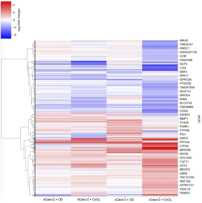
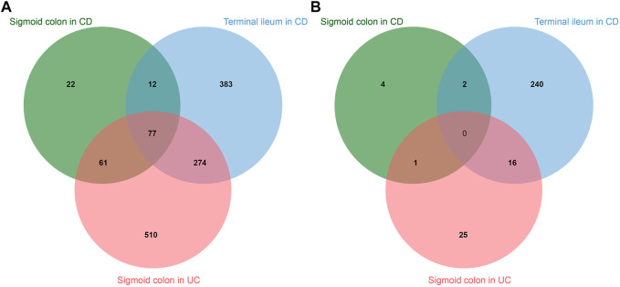
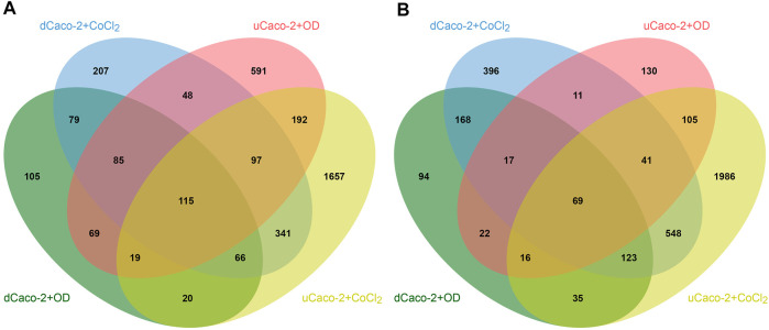
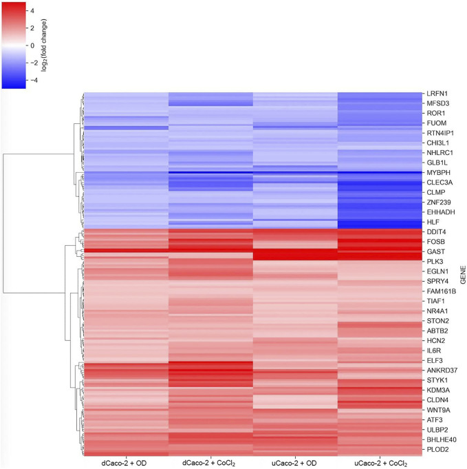
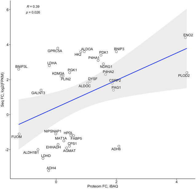
Intestinal epithelial cells exist in physiological hypoxia, leading to hypoxia-inducible factor (HIF) activation and supporting barrier function and cell metabolism of the intestinal epithelium. In contrast, pathological hypoxia is a common feature of some chronic disorders, including inflammatory bowel disease (IBD). This work was aimed at studying HIF-associated changes in the intestinal epithelium in IBD. In the first step, a list of genes responding to chemical activation of hypoxia was obtained in an intestinal cell model with RNA sequencing. Cobalt (II) chloride and oxyquinoline treatment of both undifferentiated and differentiated Caco-2 cells activate the HIF-signaling pathway according to gene set enrichment analysis. The core gene set responding to chemical hypoxia stimulation in the intestinal model included 115 upregulated and 69 downregulated genes. Of this set, protein product was detected for 32 genes, and fold changes in proteome and RNA sequencing significantly correlate. Analysis of publicly available RNA sequencing set of the intestinal epithelial cells of patients with IBD confirmed HIF-1 signaling pathway activation in sigmoid colon of patients with ulcerative colitis and terminal ileum of patients with Crohn's disease. Of the core gene set from the gut hypoxia model, expression activation of ITGA5 and PLAUR genes encoding integrin α5 and urokinase-type plasminogen activator receptor (uPAR) was detected in IBD specimens. The interaction of these molecules can activate cell migration and regenerative processes in the epithelium. Transcription factor analysis with the previously developed miRGTF tool revealed the possible role of HIF1A and NFATC1 in the regulation of ITGA5 and PLAUR gene expression. Detected genes can serve as markers of IBD progression and intestinal hypoxia.
肠上皮细胞存在于生理性缺氧环境中,可导致缺氧诱导因子(HIF)激活,并维持肠上皮的屏障功能和细胞代谢。相比之下,病理性缺氧是包括炎症性肠病(IBD)在内的一些慢性疾病的常见特征。这项研究旨在探究IBD中肠上皮细胞与HIF相关的变化。第一步,通过RNA测序在肠细胞模型中获得了对化学性缺氧激活有反应的基因列表。根据基因集富集分析,用氯化钴(II)和氧喹啉处理未分化和分化的Caco-2细胞均可激活HIF信号通路。肠道模型中对化学性缺氧刺激有反应的核心基因集包括115个上调基因和69个下调基因。在这一组基因中,检测到32个基因的蛋白质产物,蛋白质组和RNA测序中的倍数变化显著相关。对IBD患者肠上皮细胞的公开可用RNA测序数据集的分析证实,溃疡性结肠炎患者的乙状结肠和克罗恩病患者的回肠末端中HIF-1信号通路被激活。在IBD标本中检测到肠道缺氧模型核心基因集中编码整合素α5和尿激酶型纤溶酶原激活剂受体(uPAR)的ITGA5和PLAUR基因的表达激活。这些分子的相互作用可激活上皮细胞的迁移和再生过程。使用先前开发的miRGTF工具进行转录因子分析,揭示了HIF1A和NFATC1在调节ITGA5和PLAUR基因表达中的可能作用。检测到的基因可作为IBD进展和肠道缺氧的标志物。