Tremblay Christina, Rahayel Shady, Vo Andrew, Morys Filip, Shafiei Golia, Abbasi Nooshin, Markello Ross D, Gan-Or Ziv, Misic Bratislav, Dagher Alain
Montreal Neurological Institute, McGill University, Montreal, QC H3A 2B4, Canada.
Centre for Advanced Research in Sleep Medicine, Hôpital du Sacré-Cœur de Montréal, Montreal, QC H4J 1C5, Canada.
Brain Commun. 2021 Nov 17;3(4):fcab269. doi: 10.1093/braincomms/fcab269. eCollection 2021.
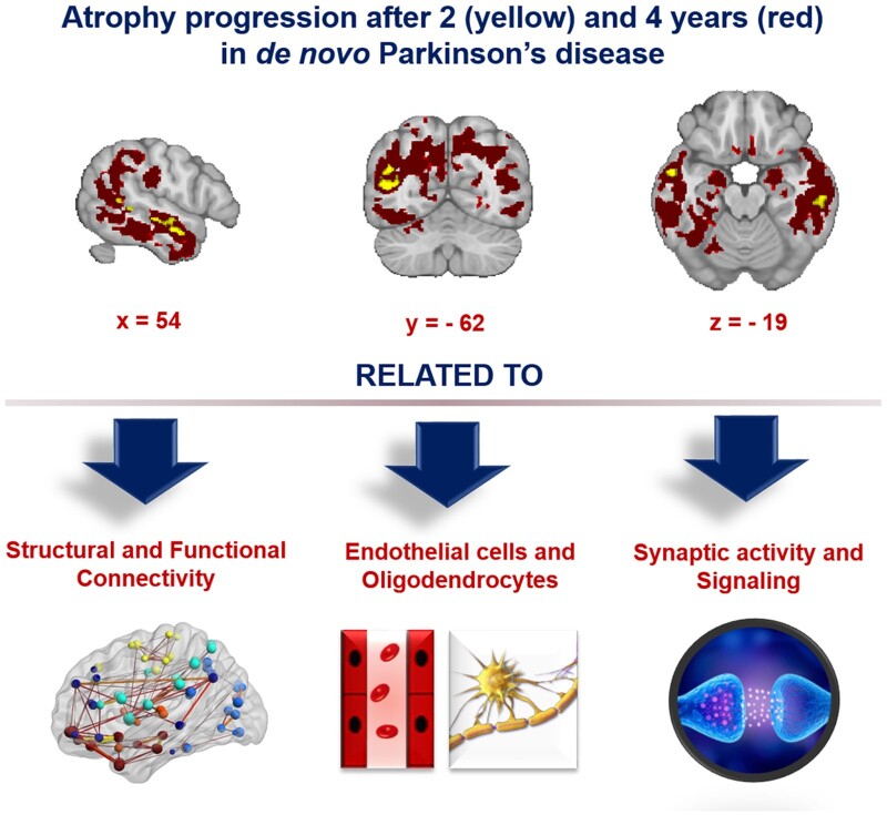
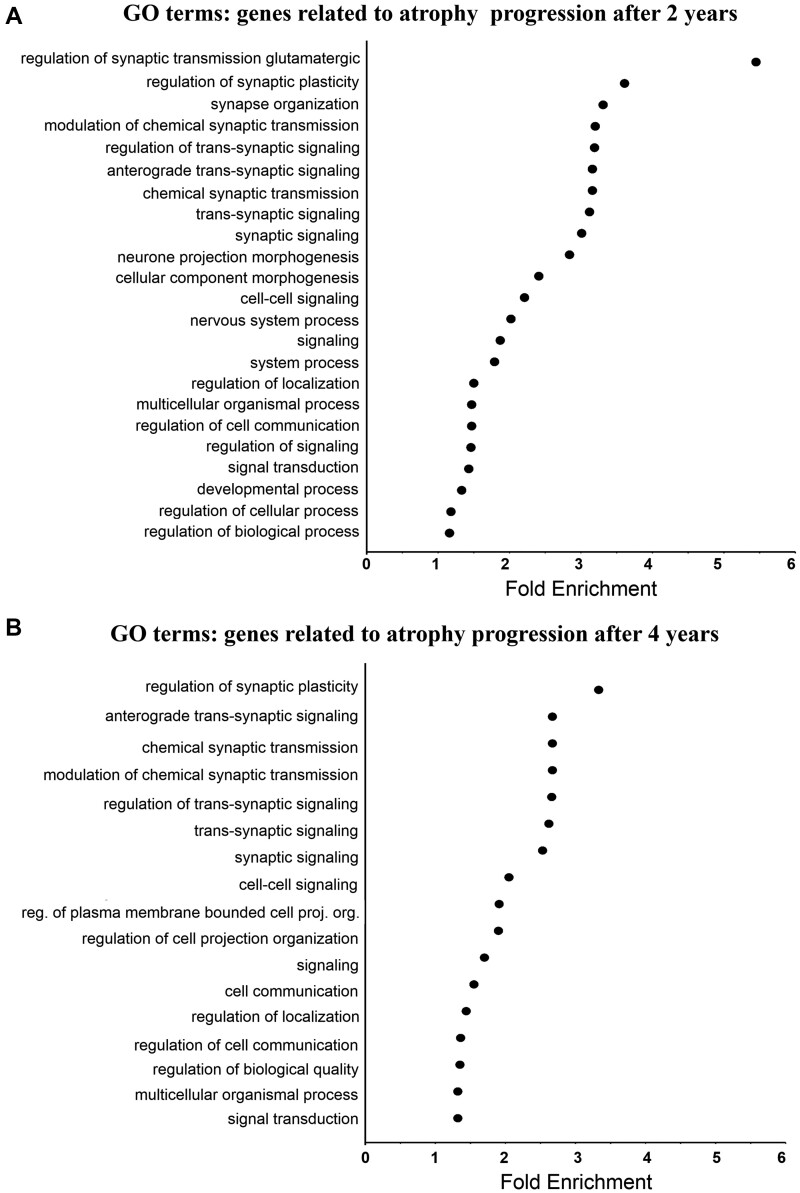
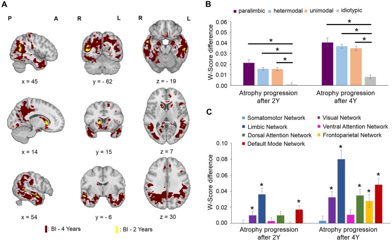
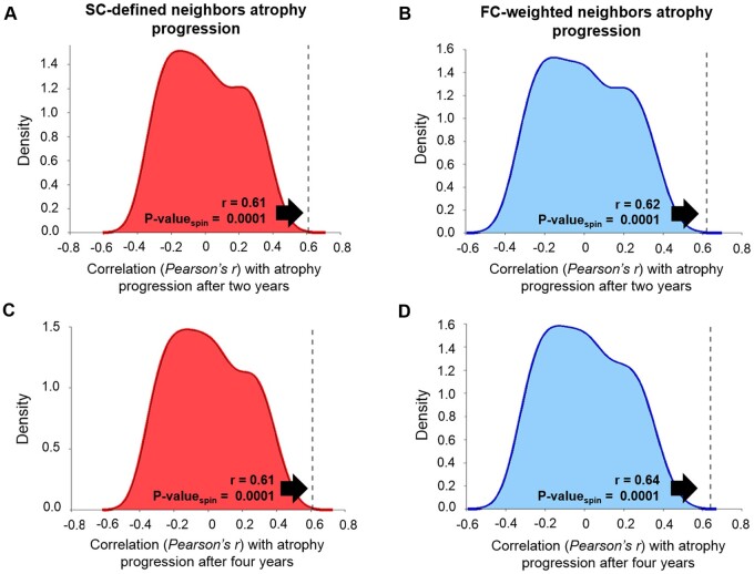
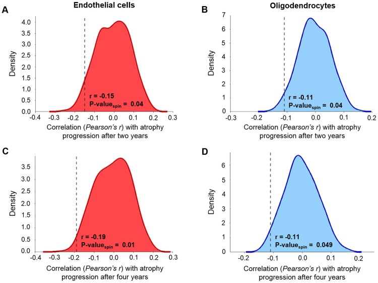
Brain atrophy has been reported in the early stages of Parkinson's disease, but there have been few longitudinal studies. How intrinsic properties of the brain, such as anatomical connectivity, local cell-type distribution and gene expression combine to determine the pattern of disease progression also remains unknown. One hypothesis proposes that the disease stems from prion-like propagation of misfolded alpha-synuclein via the connectome that might cause varying degrees of tissue damage based on local properties. Here, we used MRI data from the Parkinson Progression Markers Initiative to map the progression of brain atrophy over 1, 2 and 4 years compared with baseline. We derived atrophy maps for four time points using deformation-based morphometry applied to T-weighted MRI from 120 Parkinson's disease patients, 74 of whom had imaging at all four time points (50 Men: 24 Women) and 157 healthy control participants (115 Men: 42 Women). In order to determine factors that may influence neurodegeneration, we related atrophy progression to brain structural and functional connectivity, cell-type expression and gene ontology enrichment analyses. After regressing out the expected age and sex effects associated with normal ageing, we found that atrophy significantly progressed over 2 and 4 years in the caudate, nucleus accumbens, hippocampus and posterior cortical regions. This progression was shaped by both structural and functional brain connectivity. Also, the progression of atrophy was more pronounced in regions with a higher expression of genes related to synapses and was inversely related to the prevalence of oligodendrocytes and endothelial cells. In sum, we demonstrate that the progression of atrophy in Parkinson's disease is in line with the prion-like propagation hypothesis of alpha-synuclein and provide evidence that synapses may be especially vulnerable to synucleinopathy. In addition to identifying vulnerable brain regions, this study reveals different factors that may be implicated in the neurotoxic mechanisms leading to progression in Parkinson's disease. All brain maps generated here are available on request.
帕金森病早期已报道存在脑萎缩,但纵向研究较少。大脑的内在特性,如解剖连接性、局部细胞类型分布和基因表达如何共同决定疾病进展模式也尚不清楚。一种假说认为,该疾病源于错误折叠的α-突触核蛋白通过连接组的朊病毒样传播,这可能根据局部特性导致不同程度的组织损伤。在此,我们使用帕金森病进展标志物倡议组织的MRI数据,绘制了与基线相比1年、2年和4年期间脑萎缩的进展情况。我们对120例帕金森病患者的T加权MRI应用基于变形的形态测量法,得出了四个时间点的萎缩图谱,其中74例患者在所有四个时间点均有成像(男性50例:女性24例),以及157名健康对照参与者(男性115例:女性42例)。为了确定可能影响神经退行性变的因素,我们将萎缩进展与脑结构和功能连接性、细胞类型表达及基因本体富集分析相关联。在排除与正常衰老相关的预期年龄和性别影响后,我们发现尾状核、伏隔核、海马体和后皮质区域在2年和4年期间萎缩显著进展。这种进展受到脑结构和功能连接性的影响。此外,在与突触相关基因表达较高的区域,萎缩进展更为明显,且与少突胶质细胞和内皮细胞的患病率呈负相关。总之,我们证明帕金森病萎缩进展符合α-突触核蛋白的朊病毒样传播假说,并提供证据表明突触可能特别易受突触核蛋白病影响。除了识别易损脑区外,本研究还揭示了可能与导致帕金森病进展的神经毒性机制有关的不同因素。此处生成的所有脑图谱可应要求提供。