Department of Anatomy & Neurosciences, Amsterdam Neuroscience, Amsterdam UMC, Vrije Universiteit Amsterdam, Amsterdam, the Netherlands.
Imaging Genetics Center, Mark and Mary Stevens Neuroimaging and Informatics Institute, Keck School of Medicine, University of Southern California, Marina del Rey, California, USA.
Mov Disord. 2021 Nov;36(11):2583-2594. doi: 10.1002/mds.28706. Epub 2021 Jul 20.
Brain structure abnormalities throughout the course of Parkinson's disease have yet to be fully elucidated.
Using a multicenter approach and harmonized analysis methods, we aimed to shed light on Parkinson's disease stage-specific profiles of pathology, as suggested by in vivo neuroimaging.
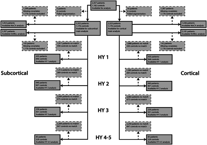
Individual brain MRI and clinical data from 2357 Parkinson's disease patients and 1182 healthy controls were collected from 19 sources. We analyzed regional cortical thickness, cortical surface area, and subcortical volume using mixed-effects models. Patients grouped according to Hoehn and Yahr stage were compared with age- and sex-matched controls. Within the patient sample, we investigated associations with Montreal Cognitive Assessment score.
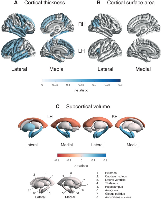
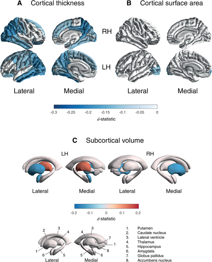
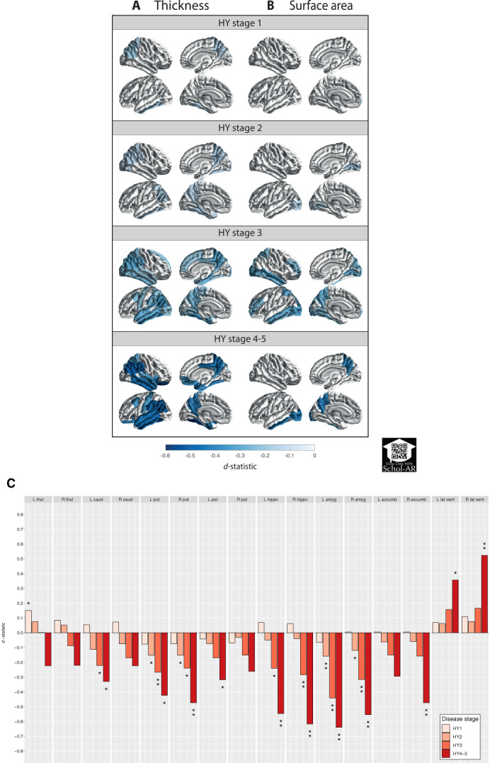
Overall, patients showed a thinner cortex in 38 of 68 regions compared with controls (d = -0.20, d = -0.09). The bilateral putamen (d = -0.14, d = -0.14) and left amygdala (d = -0.13) were smaller in patients, whereas the left thalamus was larger (d = 0.13). Analysis of staging demonstrated an initial presentation of thinner occipital, parietal, and temporal cortices, extending toward rostrally located cortical regions with increased disease severity. From stage 2 and onward, the bilateral putamen and amygdala were consistently smaller with larger differences denoting each increment. Poorer cognition was associated with widespread cortical thinning and lower volumes of core limbic structures.
Our findings offer robust and novel imaging signatures that are generally incremental across but in certain regions specific to disease stages. Our findings highlight the importance of adequately powered multicenter collaborations. © 2021 The Authors. Movement Disorders published by Wiley Periodicals LLC on behalf of International Parkinson and Movement Disorder Society.
帕金森病的大脑结构异常在整个病程中尚未得到充分阐明。
采用多中心方法和协调分析方法,我们旨在根据体内神经影像学提示,阐明帕金森病特定阶段的病理学特征。
从 19 个来源收集了 2357 名帕金森病患者和 1182 名健康对照者的个体脑 MRI 和临床数据。我们使用混合效应模型分析了区域皮质厚度、皮质表面积和皮质下体积。根据 Hoehn 和 Yahr 分期将患者分组,并与年龄和性别匹配的对照组进行比较。在患者样本中,我们研究了与蒙特利尔认知评估评分的相关性。
总体而言,与对照组相比,患者在 68 个区域中有 38 个区域的皮质较薄(d = -0.20,d = -0.09)。双侧壳核(d = -0.14,d = -0.14)和左侧杏仁核(d = -0.13)体积较小,而左侧丘脑体积较大(d = 0.13)。分期分析显示,最初表现为枕叶、顶叶和颞叶皮质变薄,随着疾病严重程度的增加,逐渐扩展到更靠近颅侧的皮质区域。从第 2 阶段开始,双侧壳核和杏仁核的体积持续减小,每个阶段的差异都更大。认知功能较差与广泛的皮质变薄和核心边缘结构的体积降低有关。
我们的发现提供了稳健且新颖的影像学特征,这些特征在整个病程中普遍呈递增趋势,但在某些区域则特定于疾病阶段。我们的研究结果强调了充分发挥多中心合作的重要性。© 2021 作者。运动障碍由 Wiley 期刊公司代表国际帕金森病和运动障碍协会出版。