Yuan Yifan, Leiby Katherine L, Greaney Allison M, Raredon Micha Sam Brickman, Qian Hong, Schupp Jonas C, Engler Alexander J, Baevova Pavlina, Adams Taylor S, Kural Mehmet H, Wang Juan, Obata Tomohiro, Yoder Mervin C, Kaminski Naftali, Niklason Laura E
Vascular Biology and Therapeutics Program, Yale University School of Medicine, New Haven, CT, United States.
Department of Anesthesiology, Yale University, New Haven, CT, United States.
Front Bioeng Biotechnol. 2021 Nov 19;9:760309. doi: 10.3389/fbioe.2021.760309. eCollection 2021.
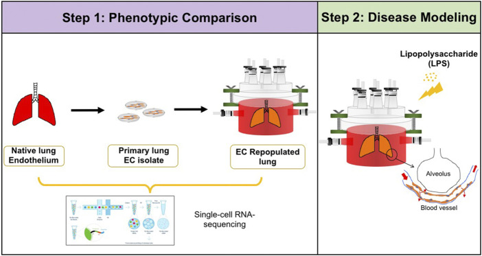
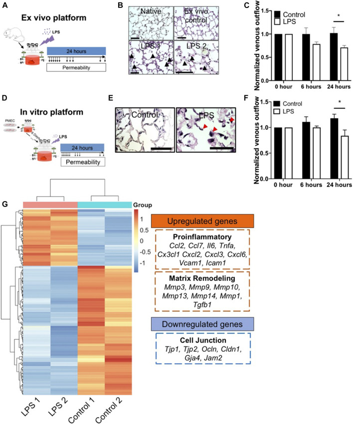
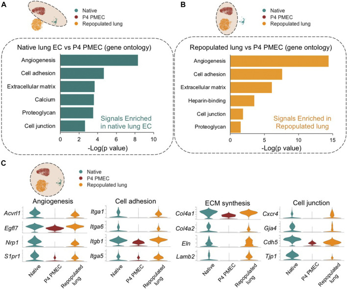
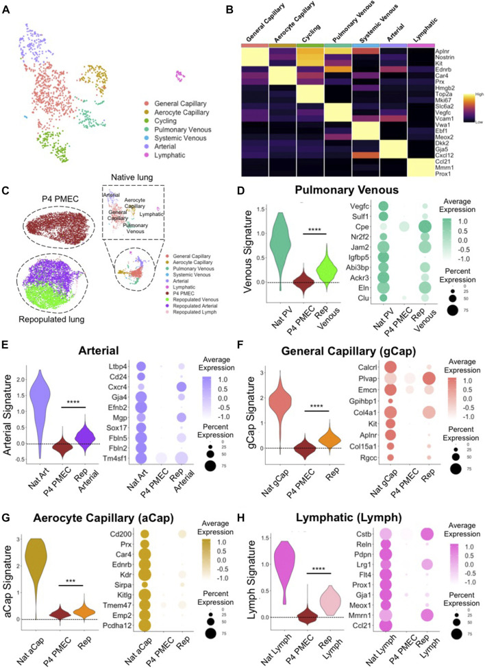
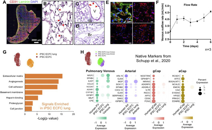
The development of an system for the study of lung vascular disease is critical to understanding human pathologies. Conventional culture systems fail to fully recapitulate native microenvironmental conditions and are typically limited in their ability to represent human pathophysiology for the study of disease and drug mechanisms. Whole organ decellularization provides a means to developing a construct that recapitulates structural, mechanical, and biological features of a complete vascular structure. Here, we developed a culture protocol to improve endothelial cell coverage in whole lung scaffolds and used single-cell RNA-sequencing analysis to explore the impact of decellularized whole lung scaffolds on endothelial phenotypes and functions in a biomimetic bioreactor system. Intriguingly, we found that the phenotype and functional signals of primary pulmonary microvascular revert back-at least partially-toward native lung endothelium. Additionally, human induced pluripotent stem cell-derived endothelium cultured in decellularized lung systems start to gain various native human endothelial phenotypes. Vascular barrier function was partially restored, while small capillaries remained patent in endothelial cell-repopulated lungs. To evaluate the ability of the engineered endothelium to modulate permeability in response to exogenous stimuli, lipopolysaccharide (LPS) was introduced into repopulated lungs to simulate acute lung injury. After LPS treatment, proinflammatory signals were significantly increased and the vascular barrier was impaired. Taken together, these results demonstrate a novel platform that recapitulates some pulmonary microvascular functions and phenotypes at a whole organ level. This development may help pave the way for using the whole organ engineering approach to model vascular diseases.
开发用于研究肺血管疾病的系统对于理解人类病理学至关重要。传统的培养系统无法完全重现天然的微环境条件,并且在代表用于疾病和药物机制研究的人类病理生理学方面通常能力有限。全器官去细胞化提供了一种开发构建体的方法,该构建体可重现完整血管结构的结构、机械和生物学特征。在这里,我们开发了一种培养方案,以提高全肺支架中的内皮细胞覆盖率,并使用单细胞RNA测序分析来探索去细胞化全肺支架对仿生生物反应器系统中内皮细胞表型和功能的影响。有趣的是,我们发现原发性肺微血管的表型和功能信号至少部分地恢复到天然肺内皮。此外,在去细胞化肺系统中培养的人诱导多能干细胞衍生的内皮开始获得各种天然的人类内皮表型。血管屏障功能部分恢复,而小毛细血管在内皮细胞重新填充的肺中保持通畅。为了评估工程化内皮细胞响应外源性刺激调节通透性的能力,将脂多糖(LPS)引入重新填充的肺中以模拟急性肺损伤。LPS处理后,促炎信号显著增加,血管屏障受损。综上所述,这些结果证明了一个在全器官水平上重现一些肺微血管功能和表型的新平台。这一进展可能有助于为使用全器官工程方法模拟血管疾病铺平道路。