Military HIV Research Program, Walter Reed Army Institute of Research, Silver Spring, Maryland, United States of America.
Henry M. Jackson Foundation for the Advancement of Military Medicine, Bethesda, Maryland, United States of America.
PLoS Pathog. 2021 Dec 7;17(12):e1010105. doi: 10.1371/journal.ppat.1010105. eCollection 2021 Dec.
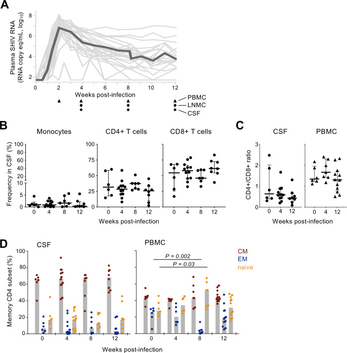
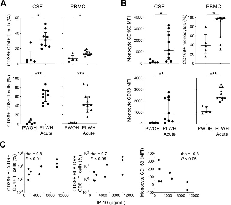
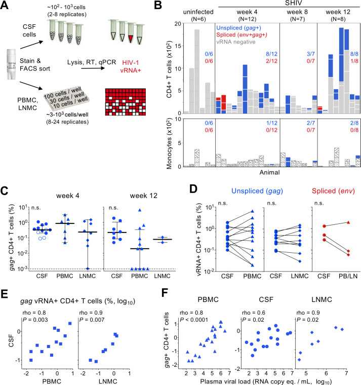
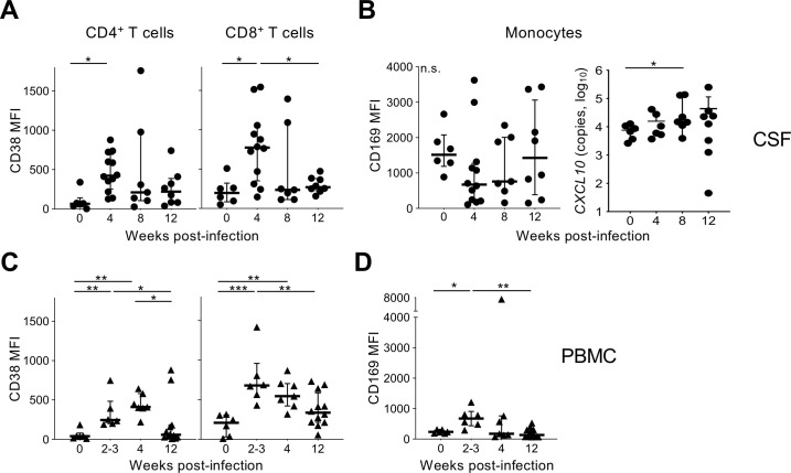
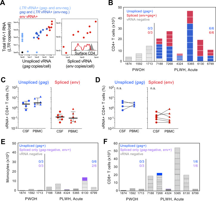
HIV-1 replication within the central nervous system (CNS) impairs neurocognitive function and has the potential to establish persistent, compartmentalized viral reservoirs. The origins of HIV-1 detected in the CNS compartment are unknown, including whether cells within the cerebrospinal fluid (CSF) produce virus. We measured viral RNA+ cells in CSF from acutely infected macaques longitudinally and people living with early stages of acute HIV-1. Active viral transcription (spliced viral RNA) was present in CSF CD4+ T cells as early as four weeks post-SHIV infection, and among all acute HIV-1 specimens (N = 6; Fiebig III/IV). Replication-inactive CD4+ T cell infection, indicated by unspliced viral RNA in the absence of spliced viral RNA, was even more prevalent, present in CSF of >50% macaques and human CSF at ~10-fold higher frequency than productive infection. Infection levels were similar between CSF and peripheral blood (and lymph nodes in macaques), indicating comparable T cell infection across these compartments. In addition, surface markers of activation were increased on CSF T cells and monocytes and correlated with CSF soluble markers of inflammation. These studies provide direct evidence of HIV-1 replication in CD4+ T cells and broad immune activation in peripheral blood and the CNS during acute infection, likely contributing to early neuroinflammation and reservoir seeding. Thus, early initiation of antiretroviral therapy may not be able to prevent establishment of CNS viral reservoirs and sources of long-term inflammation, important targets for HIV-1 cure and therapeutic strategies.
HIV-1 在中枢神经系统 (CNS) 内的复制会损害神经认知功能,并有可能建立持久的、分隔的病毒储存库。CNS 隔室中检测到的 HIV-1 的起源尚不清楚,包括脑脊液 (CSF) 中的细胞是否产生病毒。我们纵向测量了急性感染猕猴的 CSF 中的病毒 RNA+细胞,以及处于急性 HIV-1 早期阶段的人群。早在感染 SHIV 后四周,CSF CD4+T 细胞中就存在活跃的病毒转录(拼接病毒 RNA),并且在所有急性 HIV-1 标本(N=6;Fiebig III/IV)中均存在。复制非活跃的 CD4+T 细胞感染,即缺乏拼接病毒 RNA 的未拼接病毒 RNA 存在,更为普遍,在 >50%的猕猴 CSF 和人类 CSF 中存在,其频率比有复制能力的感染高约 10 倍。CSF 和外周血(以及猕猴的淋巴结)中的感染水平相似,表明这些隔室中的 T 细胞感染程度相似。此外,CSF T 细胞和单核细胞表面激活标志物增加,并与 CSF 中可溶性炎症标志物相关。这些研究提供了直接证据,表明在急性感染期间,HIV-1 在 CD4+T 细胞中复制,并广泛激活外周血和中枢神经系统中的免疫,这可能导致早期神经炎症和储存库播种。因此,早期启动抗逆转录病毒疗法可能无法预防 CNS 病毒储存库和长期炎症的建立,这是 HIV-1 治愈和治疗策略的重要目标。