Department of Nuclear Medicine, Samsung Medical Center, Sungkyunkwan University School of Medicine, 81 Irwon-ro, Gangnam-gu, Seoul, 06351, Korea.
Department of Physical and Rehabilitation Medicine, Samsung Medical Center, Sungkyunkwan University School of Medicine, Seoul, Korea.
BMC Med Imaging. 2021 Dec 8;21(1):188. doi: 10.1186/s12880-021-00713-1.
We investigated whether preoperative lymphoscintigraphy could predict the treatment response of unilateral lymphovenous anastomosis (LVA) in patients with lower extremity lymphedema.
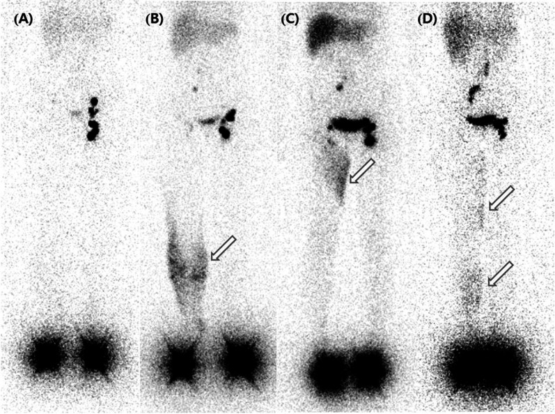
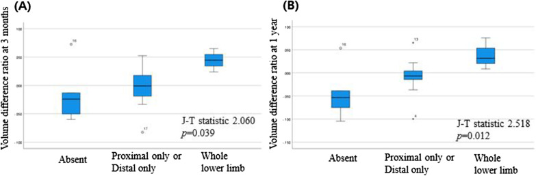
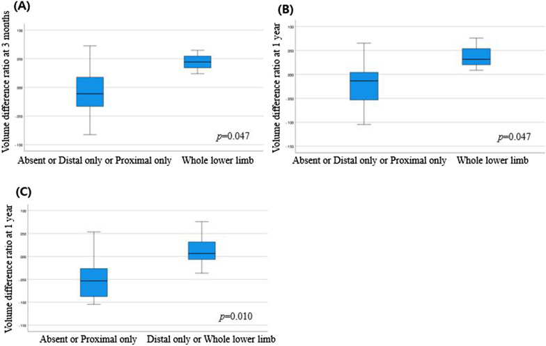
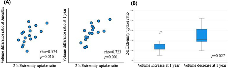
A total of 17 patients undergoing lymphoscintigraphy subsequent to LVA was included. As qualitative lymphoscintigraphic indicators, ilioinguinal lymph node uptake, main lymphatic vessel, collateral vessel, and four types of dermal backflow patterns (absent; distal only; proximal only; whole lower limb) were evaluated. Lymph node uptake ratio, extremity uptake ratio, and injection site clearance ratio were obtained as quantitative lymphoscintigraphic indicators at 1 and 2-h after injection. To evaluate therapy response, the volume difference ratio of the whole lower limb at 3 months (early response) and 1 year (late response) was measured. Volume difference ratios (continuous variable and binary variable with a cut-off value of zero) were compared according to the lymphoscintigraphic variables.
The group with whole lower limb dermal backflow had a greater volume change than the other groups (p = 0.047). The group with dermal backflow in the whole lower limb OR only in the distal part had a higher rate of volume reduction than the group with dermal backflow only in the proximal part OR absent (p = 0.050). The 2-h extremity uptake ratio was the only indicator that positively correlated with early and late volume difference ratio (p = 0.016, p = 0.001). The rate of volume decrease at 1 year was high in patients with high 2-h extremity uptake ratio (p = 0.027). As the amount of dermal backflow increases, the postoperative therapeutic effect increases (p = 0.040).
Preoperative lymphoscintigraphy is useful to predict both early and late therapy response in patients with lower extremity lymphedema undergoing LVA. Both dermal backflow pattern and extremity uptake ratio may be predictive lymphoscintigraphic indicators.
本研究旨在探讨术前淋巴闪烁显像术能否预测单侧淋巴静脉吻合术(LVA)治疗下肢淋巴水肿的疗效。
共纳入 17 例行淋巴闪烁显像术且后续行 LVA 的患者。将腹股沟淋巴结摄取、主淋巴管、侧支淋巴管和四种皮肤回流模式(无;仅远端;仅近端;整个下肢)作为定性淋巴闪烁显像指标进行评估。在注射后 1 和 2 小时,获取淋巴结摄取率、肢体摄取率和注射部位清除率作为定量淋巴闪烁显像指标。为评估治疗反应,在术后 3 个月(早期反应)和 1 年(晚期反应)测量整个下肢的体积差异比。根据淋巴闪烁显像变量比较体积差异比(连续变量和以零为截值的二分类变量)。
整个下肢皮肤回流组的体积变化大于其他组(p=0.047)。整个下肢或仅远端皮肤回流组的体积减少率高于仅近端或无皮肤回流组(p=0.050)。2 小时肢体摄取率是与早期和晚期体积差异比呈正相关的唯一指标(p=0.016,p=0.001)。2 小时肢体摄取率高的患者,1 年后的体积减少率高(p=0.027)。随着皮肤回流量的增加,术后治疗效果增加(p=0.040)。
术前淋巴闪烁显像术可预测下肢淋巴水肿患者行 LVA 后的早期和晚期治疗反应。皮肤回流模式和肢体摄取率均可能是预测性淋巴闪烁显像指标。