Annu Int Conf IEEE Eng Med Biol Soc. 2021 Nov;2021:2952-2955. doi: 10.1109/EMBC46164.2021.9630727.
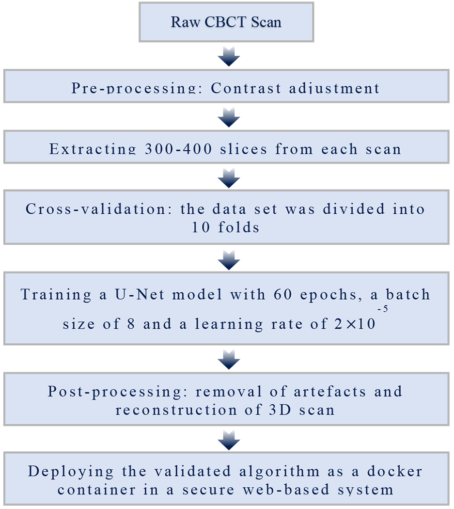
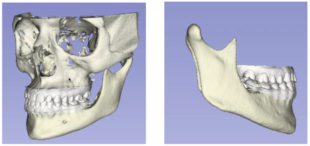
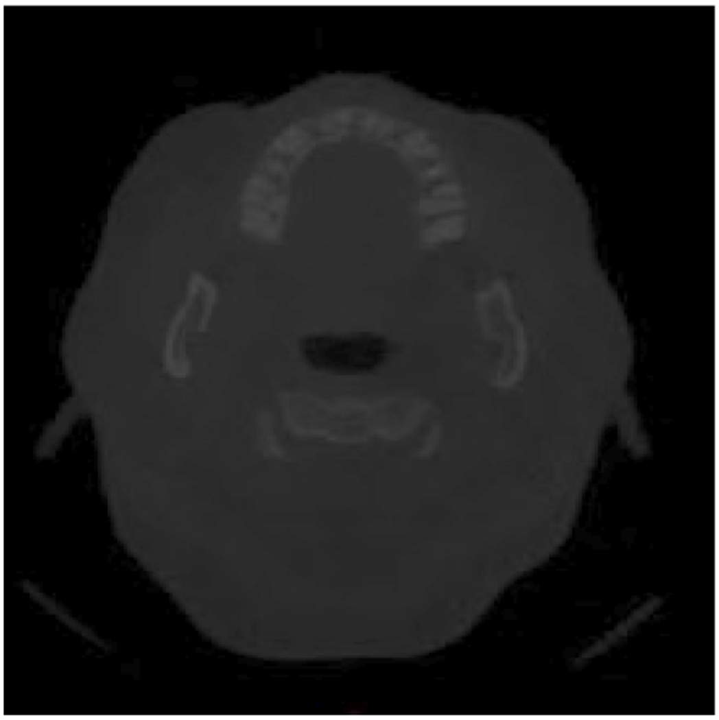
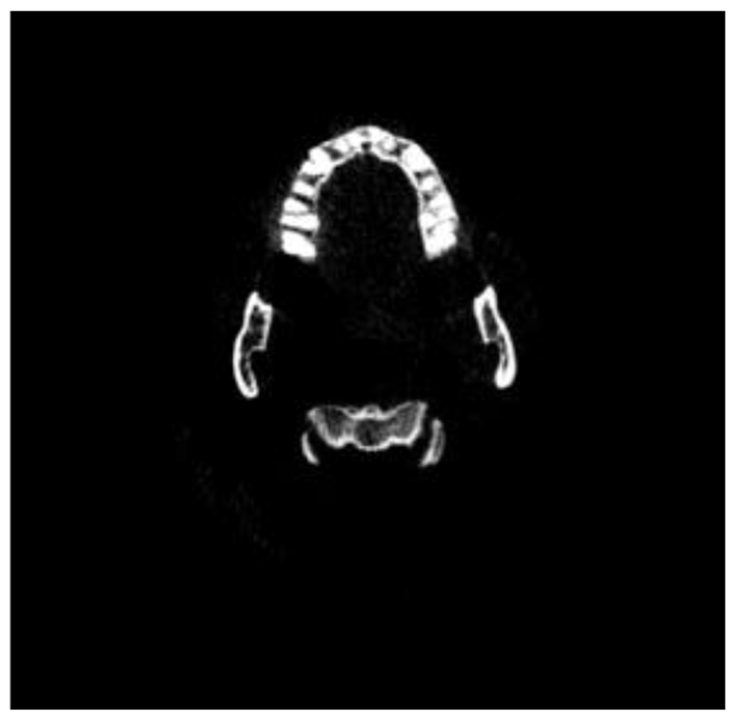
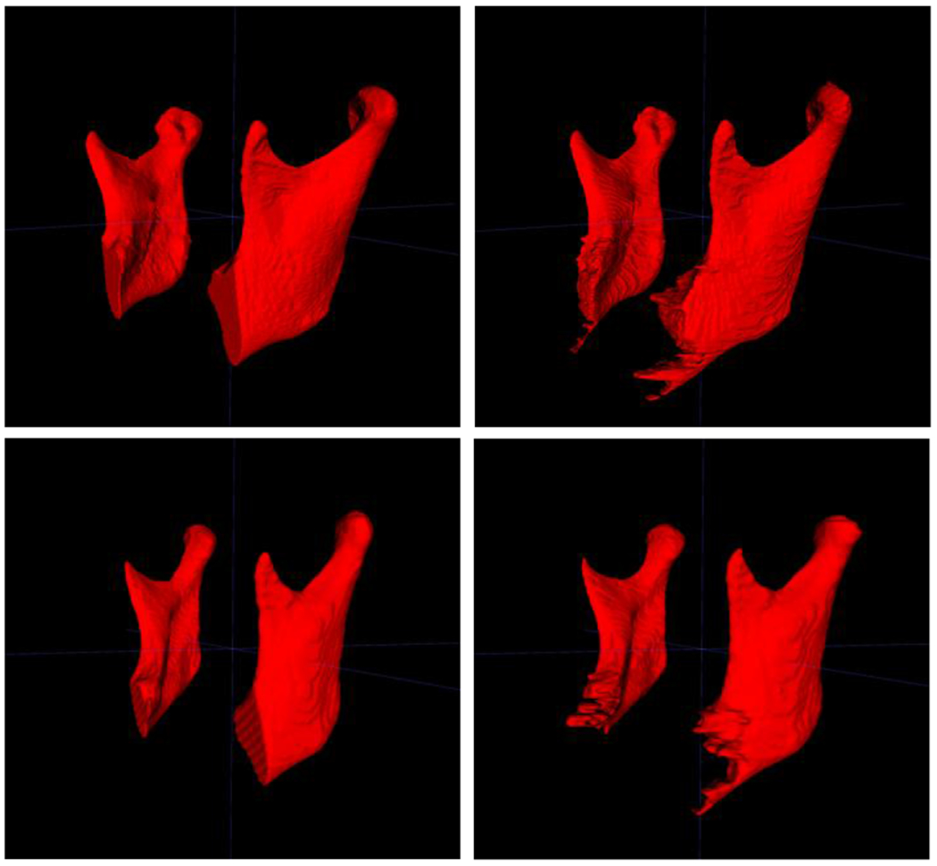
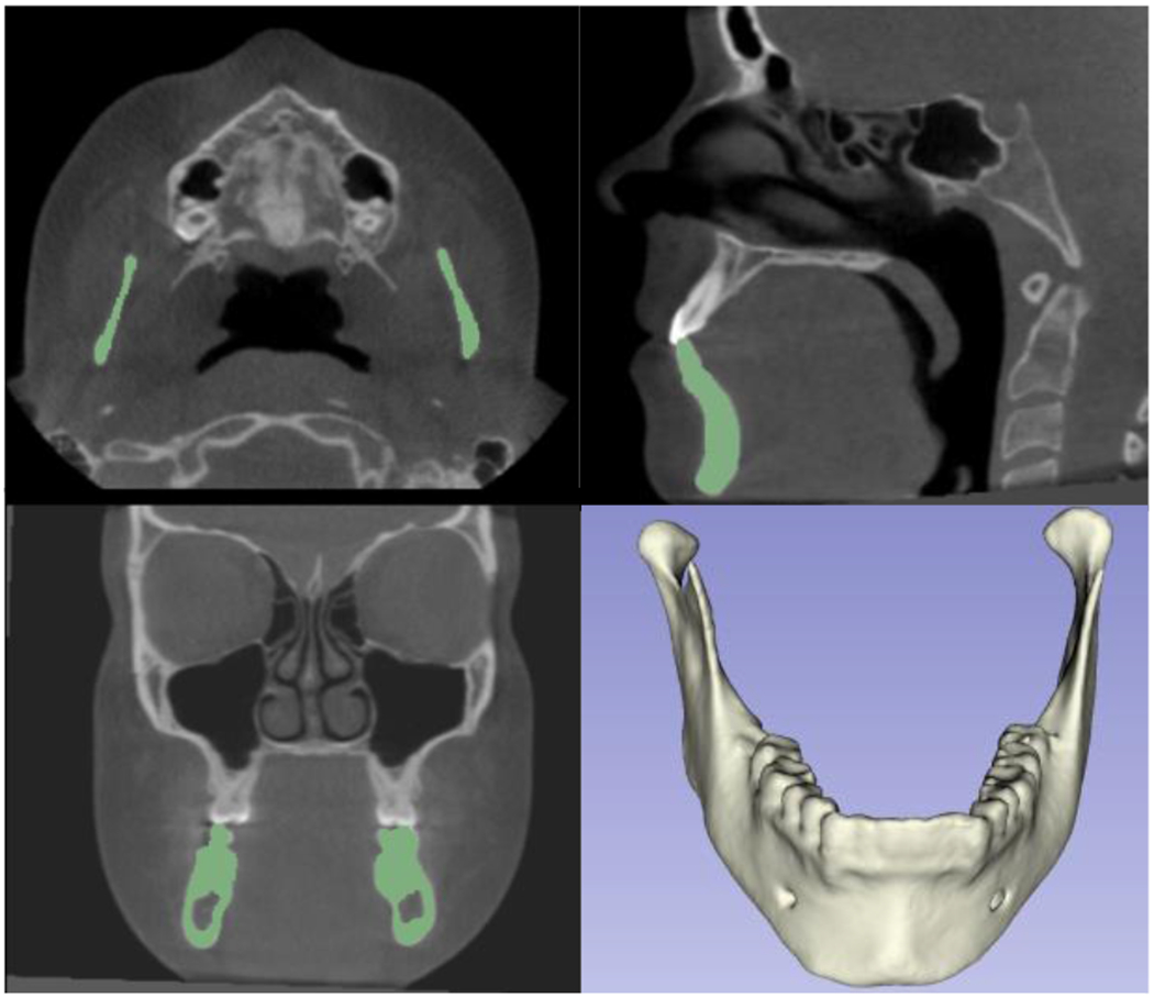
In order to diagnose TMJ pathologies, we developed and tested a novel algorithm, MandSeg, that combines image processing and machine learning approaches for automatically segmenting the mandibular condyles and ramus. A deep neural network based on the U-Net architecture was trained for this task, using 109 cone-beam computed tomography (CBCT) scans. The ground truth label maps were manually segmented by clinicians. The U-Net takes 2D slices extracted from the 3D volumetric images. All the 3D scans were cropped depending on their size in order to keep only the mandibular region of interest. The same anatomic cropping region was used for every scan in the dataset. The scans were acquired at different centers with different resolutions. Therefore, we resized all scans to 512×512 in the pre-processing step where we also performed contrast adjustment as the original scans had low contrast. After the pre-processing, around 350 slices were extracted from each scan, and used to train the U-Net model. For the cross-validation, the dataset was divided into 10 folds. The training was performed with 60 epochs, a batch size of 8 and a learning rate of 2×10. The average performance of the models on the test set presented 0.95 ± 0.05 AUC, 0.93 ± 0.06 sensitivity, 0.9998 ± 0.0001 specificity, 0.9996 ± 0.0003 accuracy, and 0.91 ± 0.03 F1 score. This study findings suggest that fast and efficient CBCT image segmentation of the mandibular condyles and ramus from different clinical data sets and centers can be analyzed effectively. Future studies can now extract radiomic and imaging features as potentially relevant objective diagnostic criteria for TMJ pathologies, such as osteoarthritis (OA). The proposed segmentation will allow large datasets to be analyzed more efficiently for disease classification.
为了诊断 TMJ 病变,我们开发并测试了一种新的算法 MandSeg,该算法结合了图像处理和机器学习方法,用于自动分割下颌骨髁突和升支。为此任务,使用 109 个锥形束 CT(CBCT)扫描对基于 U-Net 架构的深度神经网络进行了训练。通过临床医生手动分割得到地面真实标签图。U-Net 从 3D 容积图像中提取 2D 切片。所有 3D 扫描都根据其大小进行裁剪,以仅保留感兴趣的下颌区域。在数据集的每个扫描中都使用相同的解剖裁剪区域。扫描是在不同的中心以不同的分辨率采集的。因此,我们在预处理步骤中将所有扫描调整为 512×512,在该步骤中还进行了对比度调整,因为原始扫描对比度较低。预处理后,从每个扫描中提取约 350 个切片,并用于训练 U-Net 模型。对于交叉验证,将数据集分为 10 折。训练使用 60 个 epoch、批量大小为 8 和学习率为 2×10 进行。模型在测试集上的平均性能为 0.95±0.05 AUC、0.93±0.06 灵敏度、0.9998±0.0001 特异性、0.9996±0.0003 准确性和 0.91±0.03 F1 分数。本研究结果表明,可以有效地分析来自不同临床数据集和中心的快速高效的 CBCT 下颌骨髁突和升支图像分割。未来的研究现在可以提取放射组学和成像特征作为 TMJ 病变的潜在相关客观诊断标准,例如骨关节炎(OA)。拟议的分割将允许更有效地分析大型数据集以进行疾病分类。