Department of Oral and Maxillofacial Surgery, Radboud University Medical Centre, 6500 HB, P.O. Box 9101, Nijmegen, 590, the Netherlands.
Radboudumc 3DLab, Radboud University Medical Centre, Nijmegen, the Netherlands.
Clin Oral Investig. 2024 Sep 4;28(9):512. doi: 10.1007/s00784-024-05895-w.
In orthognatic surgery, one of the primary determinants for reliable three-dimensional virtual surgery planning (3D VSP) and an accurate transfer of 3D VSP to the patient in the operation room is the condylar seating. Incorrectly seated condyles would primarily affect the accuracy of maxillary-first bimaxillary osteotomies as the maxillary repositioning is dependent on the positioning of the mandible in the cone-beam computed tomography (CBCT) scan. This study aimed to develop and validate a novel tool by utilizing a deep learning algorithm that automatically evaluates the condylar seating based on CBCT images as a proof of concept.
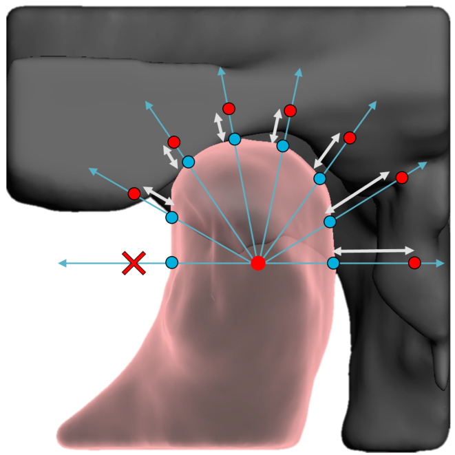
As a reference, 60 CBCT scans (120 condyles) were labeled. The automatic assessment of condylar seating included three main parts: segmentation module, ray-casting, and feed-forward neural network (FFNN). The AI-based algorithm was trained and tested using fivefold cross validation. The method's performance was evaluated by comparing the labeled ground truth with the model predictions on the validation dataset.
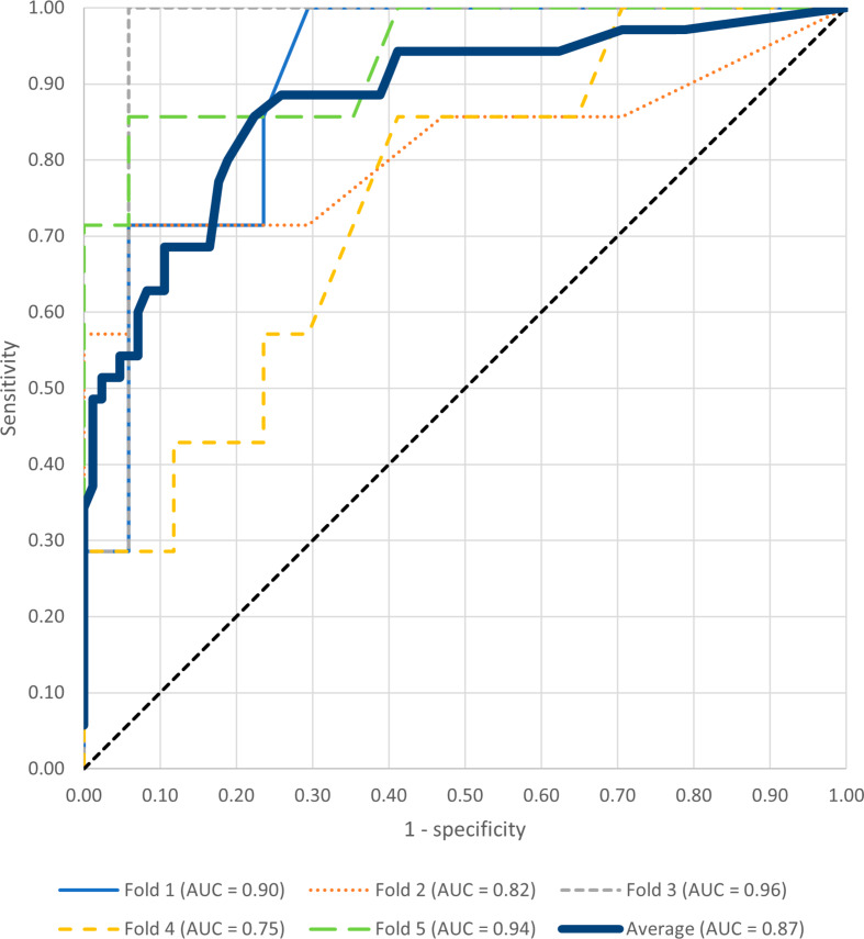
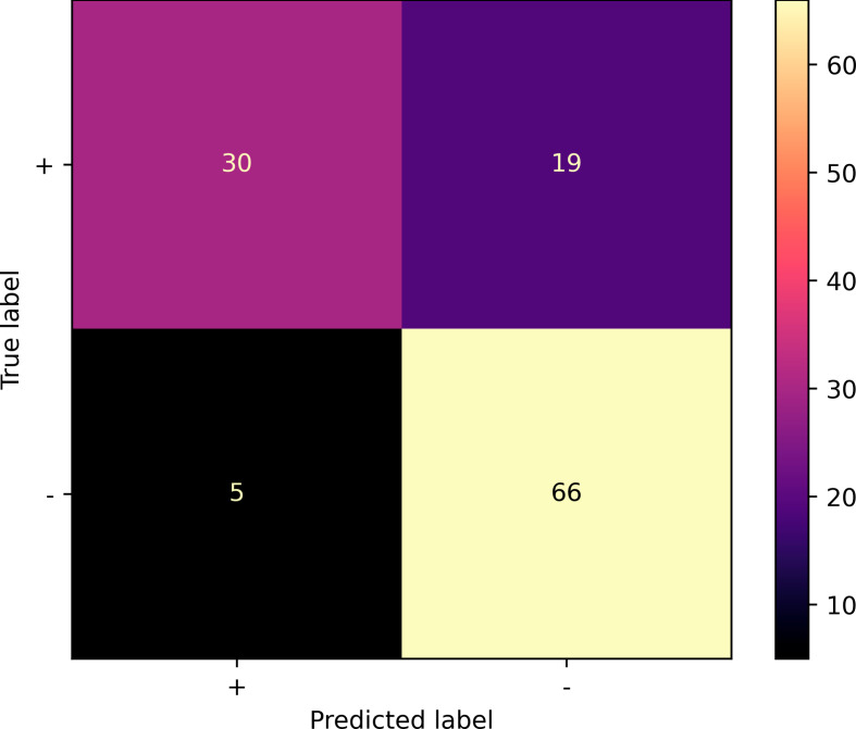
The model achieved an accuracy of 0.80, positive predictive value of 0.61, negative predictive value of 0.9 and F1-score of 0.71. The sensitivity and specificity of the model was 0.86 and 0.78, respectively. The mean AUC over all folds was 0.87.
The innovative integration of multi-step segmentation, ray-casting and a FFNN demonstrated to be a viable approach for automating condylar seating assessment and have obtained encouraging results.
Automated condylar seating assessment using deep learning may improve orthognathic surgery, preventing errors and enhancing patient outcomes in maxillary-first bimaxillary osteotomies.
在正颌手术中,可靠的三维虚拟手术规划(3D VSP)和将 3D VSP 准确转移到手术室患者的主要决定因素之一是髁突的定位。不正确定位的髁突主要会影响上颌骨双颌骨截骨术的准确性,因为上颌骨的重新定位取决于 CBCT 扫描中下颌骨的定位。本研究旨在开发和验证一种新工具,该工具利用深度学习算法根据 CBCT 图像自动评估髁突的定位,作为概念验证。
作为参考,标记了 60 个 CBCT 扫描(120 个髁突)。髁突定位的自动评估包括三个主要部分:分割模块、射线投射和前馈神经网络(FFNN)。使用五重交叉验证对基于 AI 的算法进行了训练和测试。该方法的性能通过将标记的地面实况与验证数据集上的模型预测进行比较来评估。
该模型的准确率为 0.80,阳性预测值为 0.61,阴性预测值为 0.9,F1 得分为 0.71。该模型的灵敏度和特异性分别为 0.86 和 0.78。所有折叠的平均 AUC 为 0.87。
多步分割、射线投射和 FFNN 的创新集成被证明是一种可行的方法,可用于自动评估髁突的定位,并取得了令人鼓舞的结果。
使用深度学习进行自动髁突定位评估可能会改善正颌手术,防止上颌骨双颌骨截骨术中的错误并改善患者的预后。