Stroke Center & Clinical Trial and Research Center for Stroke, Department of Neurology, The First Hospital of Jilin University, Changchun, China.
China National Comprehensive Stroke Center, Changchun, China.
CNS Neurosci Ther. 2022 Feb;28(2):298-306. doi: 10.1111/cns.13778. Epub 2021 Dec 11.
Cerebral small vessel disease (CSVD) is characterized by functional and structural changes in small vessels. We aimed to elucidate the relationship between dynamic cerebral autoregulation (dCA) and neuroimaging characteristics of CSVD.
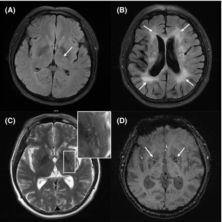
A case-control study was performed. Cerebral blood flow velocity (CBFV) of bilateral middle cerebral arteries and spontaneous arterial blood pressure were simultaneously recorded. Transfer function analysis was used to calculate dCA parameters (phase, gain, and the rate of recovery of CBFV [RoRc]). Neuroimaging characteristics of CSVD patients were evaluated, including lacunes, white matter hyperintensities (WMH), cerebral microbleeds (CMBs), perivascular spaces (PVS), and the total CSVD burden.
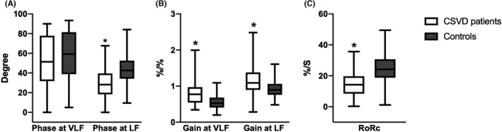
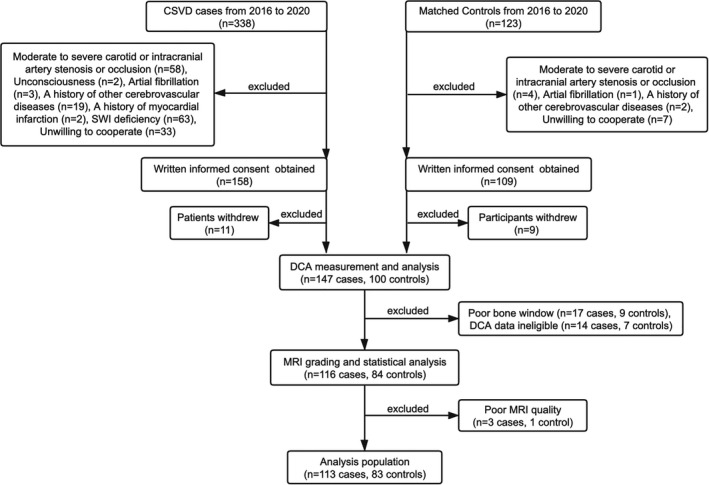
Overall, 113 patients and 83 controls were enrolled. Compared with the control group, the phase at low frequency and the RoRc in CSVD patients were lower, and the gain at very low and low frequencies were higher, indicating bilaterally impaired dCA. Total CSVD burden, WMH (total, periventricular and deep), severe PVS, and lobar CMBs were independently correlated with the phase at low frequency.
Our findings suggested that dCA was compromised in CSVD patients, and some specific neuroimaging characteristics (the total CSVD burden, WMH, severe PVS and lobar CMBs) might indicate more severe dCA impairment in CSVD patients.
脑小血管病(CSVD)的特征是小血管的功能和结构改变。我们旨在阐明动态脑自动调节(dCA)与 CSVD 的神经影像学特征之间的关系。
进行了一项病例对照研究。同时记录双侧大脑中动脉的脑血流速度(CBFV)和自发性动脉血压。使用传递函数分析来计算 dCA 参数(相位、增益和 CBFV 的恢复率[RoRc])。评估 CSVD 患者的神经影像学特征,包括腔隙、脑白质高信号(WMH)、脑微出血(CMBs)、血管周围间隙(PVS)和总 CSVD 负担。
共有 113 例患者和 83 例对照纳入研究。与对照组相比,CSVD 患者的低频相位和 RoRc 较低,极低和低频的增益较高,表明双侧 dCA 受损。总 CSVD 负担、WMH(总、脑室周围和深部)、严重 PVS 和皮质下 CMBs 与低频相位独立相关。
我们的研究结果表明,CSVD 患者的 dCA 受损,一些特定的神经影像学特征(总 CSVD 负担、WMH、严重 PVS 和皮质下 CMBs)可能表明 CSVD 患者的 dCA 损害更严重。