Mohr Hermine, Foscarini Alessia, Steiger Katja, Ballke Simone, Rischpler Christoph, Schilling Franz, Pellegata Natalia S
Institute for Diabetes and Cancer, Helmholtz Zentrum München, Ingolstaedter Landstrasse 1, 85764, Neuherberg, Germany.
Joint Heidelberg-IDC Translational Diabetes Program, Heidelberg University Hospital, Heidelberg, Germany.
EJNMMI Res. 2021 Dec 11;11(1):121. doi: 10.1186/s13550-021-00855-x.
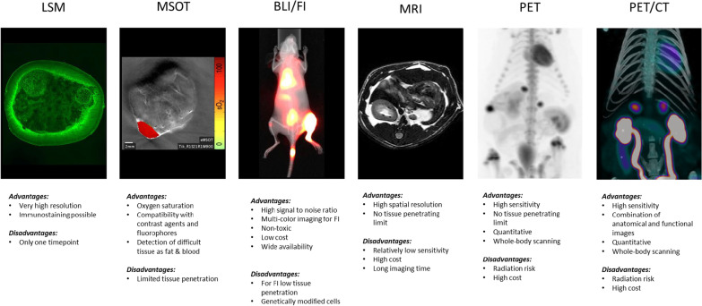
Pheochromocytomas (PCCs) and paragangliomas (PGLs), together referred to as PPGLs, are rare chromaffin cell-derived tumors. They require timely diagnosis as this is the only way to achieve a cure through surgery and because of the potentially serious cardiovascular complications and sometimes life-threatening comorbidities that can occur if left untreated. The biochemical diagnosis of PPGLs has improved over the last decades, and the knowledge of the underlying genetics has dramatically increased. In addition to conventional anatomical imaging by CT and MRI for PPGL detection, new functional imaging modalities have emerged as very useful for patient surveillance and stratification for therapy. The availability of validated and predictive animal models of cancer is essential for translating molecular, imaging and therapy response findings from the bench to the bedside. This is especially true for rare tumors, such as PPGLs, for which access to large cohorts of patients is limited. There are few animal models of PPGLs that have been instrumental in refining imaging modalities for early tumor detection, as well as in identifying and evaluating novel imaging tracers holding promise for the detection and/or treatment of human PPGLs. The in vivo PPGL models mainly include xenografts/allografts generated by engrafting rat or mouse cell lines, as no representative human cell line is available. In addition, there is a model of endogenous PCCs (i.e., MENX rats) that was characterized in our laboratory. In this review, we will summarize the contribution that various representative models of PPGL have given to the visualization of these tumors in vivo and we present an example of a tracer first evaluated in MENX rats, and then translated to the detection of these tumors in human patients. In addition, we will illustrate briefly the potential of ex vivo biological imaging of intact adrenal glands in MENX rats.
嗜铬细胞瘤(PCC)和副神经节瘤(PGL)统称为PPGL,是罕见的嗜铬细胞源性肿瘤。它们需要及时诊断,因为这是通过手术实现治愈的唯一途径,而且如果不治疗,可能会出现严重的心血管并发症,有时还会出现危及生命的合并症。在过去几十年中,PPGL的生化诊断有所改善,对其潜在遗传学的了解也大幅增加。除了通过CT和MRI进行传统的解剖学成像以检测PPGL外,新的功能成像模式已成为对患者进行监测和治疗分层的非常有用的手段。经过验证的癌症预测动物模型对于将分子、成像和治疗反应研究结果从实验室转化到临床至关重要。对于罕见肿瘤,如PPGL,情况尤其如此,因为获得大量患者队列的机会有限。很少有PPGL动物模型有助于改进早期肿瘤检测的成像模式,以及识别和评估有望用于检测和/或治疗人类PPGL的新型成像示踪剂。体内PPGL模型主要包括通过移植大鼠或小鼠细胞系产生的异种移植/同种异体移植,因为没有代表性的人类细胞系可用。此外,我们实验室还建立了一种内源性PCC模型(即MENX大鼠)。在本综述中,我们将总结各种代表性PPGL模型对这些肿瘤体内可视化的贡献,并介绍一种首先在MENX大鼠中评估,然后转化用于检测人类患者这些肿瘤的示踪剂的例子。此外,我们将简要说明MENX大鼠完整肾上腺的体外生物成像潜力。