Department of Ophthalmology, Keelung Chang Gung Memorial Hospital, Keelung, Taiwan.
Department of Ophthalmology, Jen-Ai Hospital, Taichung, Taiwan.
Transl Vis Sci Technol. 2021 Dec 1;10(14):9. doi: 10.1167/tvst.10.14.9.
To investigate the association between retinal neurovascular biomarkers and early cognitive impairment among patients with chronic kidney disease (CKD).
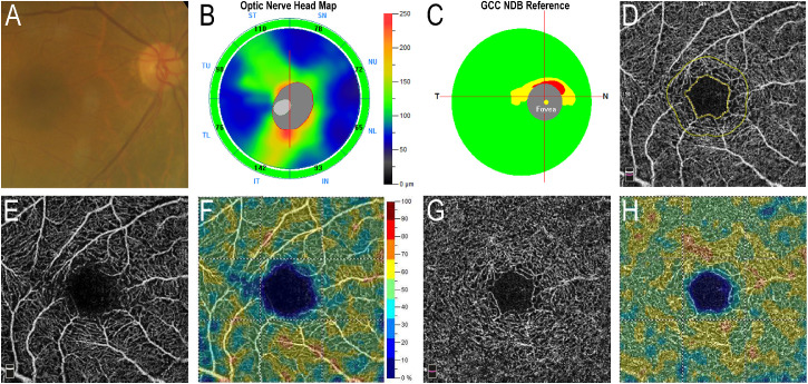
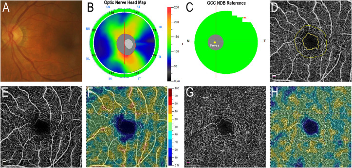
Patients with CKD stage ≥3 were evaluated using the standardized Mini-Mental State Examination (MMSE). Patients were classified as having a low (<24), middle (24 to 27), and high (>27) MMSE level. Retinal nerve fiber layer thickness, ganglion cell complex (GCC) thickness, GCC global loss volume, and GCC focal loss volume were measured using optical coherence tomography (OCT). Superficial vascular plexus vessel density, deep vascular plexus vessel density (DVP-VD), and size of the foveal avascular zone were obtained by OCT angiography.
The study enrolled 177 patients with a mean ± SD age of 64.7 ± 6.6 years. The mean ± SD MMSE score was 27.25 ± 2.30. Thirteen, 65, and 99 patients were classified as having a low, middle, and high MMSE level, respectively. The patients with a high MMSE level were younger, had more years of education, had less severe CKD, and had higher DVP-VD than patients with a low MMSE level. The multivariable regression revealed that age (coefficient, 0.294; 95% confidence interval [CI], 0.195-0.393; P = 0.041), years of education (coefficient, 0.294; 95% CI, 0.195-0.393; P < 0.001), estimated glomerular filtration rate (coefficient, 0.019; 95% CI, 0.004-0.035; P = 0.016), and DVP-VD (coefficient, 0.109; 95% CI, 0.007-0.212; P = 0.037) were independent factors associated with MMSE score.
Retinal DVP-VD was associated with early cognitive impairment among patients with CKD.
DVP-VD measured by OCT angiography may facilitate early detection of cognitive impairment.
探讨慢性肾脏病(CKD)患者视网膜神经血管生物标志物与早期认知障碍的关系。
对 CKD 分期≥3 的患者进行标准化的简易精神状态检查(MMSE)评估。患者被分为低(<24)、中(24-27)和高(>27)MMSE 水平。使用光学相干断层扫描(OCT)测量视网膜神经纤维层厚度、节细胞复合体(GCC)厚度、GCC 整体丢失体积和 GCC 局灶性丢失体积。通过 OCT 血管造影获得浅层血管丛血管密度、深层血管丛血管密度(DVP-VD)和黄斑无血管区大小。
这项研究纳入了 177 名平均年龄为 64.7±6.6 岁的患者。平均 MMSE 评分为 27.25±2.30。13、65 和 99 名患者分别被归类为低、中、高 MMSE 水平。MMSE 水平较高的患者年龄较小、受教育年限较长、CKD 程度较轻、DVP-VD 较高。多变量回归显示,年龄(系数,0.294;95%置信区间[CI],0.195-0.393;P=0.041)、受教育年限(系数,0.294;95%CI,0.195-0.393;P<0.001)、估算肾小球滤过率(系数,0.019;95%CI,0.004-0.035;P=0.016)和 DVP-VD(系数,0.109;95%CI,0.007-0.212;P=0.037)是与 MMSE 评分相关的独立因素。
视网膜 DVP-VD 与 CKD 患者的早期认知障碍有关。
李洁