• 文献检索
  • 文档翻译
  • 深度研究
  • 学术资讯
  • Suppr Zotero 插件Zotero 插件
  • 邀请有礼
  • 套餐&价格
  • 历史记录
应用&插件
Suppr Zotero 插件Zotero 插件浏览器插件Mac 客户端Windows 客户端微信小程序
定价
高级版会员购买积分包购买API积分包
服务
文献检索文档翻译深度研究API 文档MCP 服务
关于我们
关于 Suppr公司介绍联系我们用户协议隐私条款
关注我们

Suppr 超能文献

核心技术专利:CN118964589B侵权必究
粤ICP备2023148730 号-1Suppr @ 2026

文献检索

告别复杂PubMed语法,用中文像聊天一样搜索,搜遍4000万医学文献。AI智能推荐,让科研检索更轻松。

立即免费搜索

文件翻译

保留排版,准确专业,支持PDF/Word/PPT等文件格式,支持 12+语言互译。

免费翻译文档

深度研究

AI帮你快速写综述,25分钟生成高质量综述,智能提取关键信息,辅助科研写作。

立即免费体验

脉络膜渗漏在中心性浆液性脉络膜视网膜病变中的表现。

CILIOCHOROIDAL EFFUSION IN CENTRAL SEROUS CHORIORETINOPATHY.

机构信息

Department of Ophthalmology, Graduate School of Medicine, University of the Ryukyus, Okinawa, Japan .

出版信息

Retina. 2022 Apr 1;42(4):730-737. doi: 10.1097/IAE.0000000000003376.

DOI:10.1097/IAE.0000000000003376
PMID:34907128
原文链接:https://pmc.ncbi.nlm.nih.gov/articles/PMC8946592/
Abstract

PURPOSE

To investigate the prevalence of ciliochoroidal effusion (CE) in central serous chorioretinopathy (CSC) using anterior-segment optical coherence tomography and its association with the clinical features of CSC.

METHODS

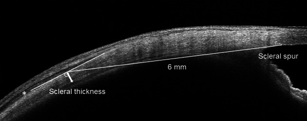
Overall, 164 eyes of 164 patients with CSC and 51 eyes of 51 age- and sex-matched normal control participants were retrospectively examined. Anterior-segment optical coherence tomography was used to assess patients with CSC and control subjects for CE and scleral thickness. Central serous chorioretinopathy eyes were divided into two groups: eyes with CE (CE group) and eyes without CE (non-CE group). Scleral thickness was measured at the point that was 6 mm posterior to the scleral spur in four directions.

RESULTS

Among the 164 eyes with CSC, 32 eyes (19.5%) displayed CE, and this proportion was significantly higher than that in control subjects (2.0%) (P = 0.001). Scleral thickness was significantly greater in the CE group compared with the non-CE group at all four directions (P < 0.05 for all). Multivariable analysis revealed that the mean scleral thickness (odds ratio: 1.01; 95% confidence interval: 1.00-1.02; P = 0.007) was significantly associated with the incidence of CE.

CONCLUSION

Central serous chorioretinopathy may accompany fluid accumulation in the anterior segment more frequently than previously expected in association with thick sclera.

摘要

目的

使用眼前节光学相干断层扫描(OCT)研究中心性浆液性脉络膜视网膜病变(CSC)中睫状体脉络膜渗漏(CE)的患病率,并探讨其与 CSC 临床特征的关系。

方法

回顾性分析了 164 例(164 只眼)CSC 患者和 51 例(51 只眼)年龄和性别匹配的正常对照组患者的资料。使用眼前节 OCT 评估 CSC 患者和对照组患者的 CE 和巩膜厚度。将 CSC 眼分为两组:有 CE(CE 组)和无 CE(非 CE 组)。在四个方向上,在巩膜突后 6mm 处测量巩膜厚度。

结果

在 164 只 CSC 眼中,32 只(19.5%)出现 CE,这一比例明显高于对照组(2.0%)(P=0.001)。CE 组的巩膜厚度在四个方向上均显著大于非 CE 组(P<0.05)。多变量分析显示,平均巩膜厚度(优势比:1.01;95%置信区间:1.00-1.02;P=0.007)与 CE 的发生显著相关。

结论

CSC 可能比以前预期的更常伴有前节液体积聚,同时伴有巩膜增厚。

https://cdn.ncbi.nlm.nih.gov/pmc/blobs/e6b7/8946592/8cf44b3bbfcf/retina-42-730-g003.jpg
https://cdn.ncbi.nlm.nih.gov/pmc/blobs/e6b7/8946592/c6736aa33c74/retina-42-730-g001.jpg
https://cdn.ncbi.nlm.nih.gov/pmc/blobs/e6b7/8946592/1b0907a2f840/retina-42-730-g002.jpg
https://cdn.ncbi.nlm.nih.gov/pmc/blobs/e6b7/8946592/8cf44b3bbfcf/retina-42-730-g003.jpg
https://cdn.ncbi.nlm.nih.gov/pmc/blobs/e6b7/8946592/c6736aa33c74/retina-42-730-g001.jpg
https://cdn.ncbi.nlm.nih.gov/pmc/blobs/e6b7/8946592/1b0907a2f840/retina-42-730-g002.jpg
https://cdn.ncbi.nlm.nih.gov/pmc/blobs/e6b7/8946592/8cf44b3bbfcf/retina-42-730-g003.jpg

相似文献

1
CILIOCHOROIDAL EFFUSION IN CENTRAL SEROUS CHORIORETINOPATHY.脉络膜渗漏在中心性浆液性脉络膜视网膜病变中的表现。
Retina. 2022 Apr 1;42(4):730-737. doi: 10.1097/IAE.0000000000003376.
2
SCLERAL THICKNESS IN THE FELLOW EYES OF PATIENTS WITH UNILATERAL CENTRAL SEROUS CHORIORETINOPATHY.单侧中心性浆液性脉络膜视网膜病变患者对侧眼巩膜厚度。
Retina. 2023 Sep 1;43(9):1573-1578. doi: 10.1097/IAE.0000000000003850.
3
Scleral Thickness in Central Serous Chorioretinopathy.脉络膜视网膜病变中心性浆液性。
Ophthalmol Retina. 2021 Mar;5(3):285-291. doi: 10.1016/j.oret.2020.07.011. Epub 2020 Jul 16.
4
Scleral Thickness in Simple Versus Complex Central Serous Chorioretinopathy.单纯型与复合型中心性浆液性脉络膜视网膜病变的巩膜厚度比较。
Am J Ophthalmol. 2024 May;261:103-111. doi: 10.1016/j.ajo.2024.01.025. Epub 2024 Jan 26.
5
Relationship Between Scleral Thickness and Choroidal Structure in Central Serous Chorioretinopathy.巩膜厚度与中心性浆液性脉络膜视网膜病变脉络膜结构的关系。
Invest Ophthalmol Vis Sci. 2023 Jan 3;64(1):16. doi: 10.1167/iovs.64.1.16.
6
Clinical Factors Related to Loculation of Fluid in Central Serous Chorioretinopathy.与中心性浆液性脉络膜视网膜病变液体积聚相关的临床因素。
Am J Ophthalmol. 2022 Mar;235:197-203. doi: 10.1016/j.ajo.2021.09.009. Epub 2021 Sep 20.
7
Assessment of the anterior scleral thickness in central serous chorioretinopathy patients by optical coherence tomography.光学相干断层扫描评估中心性浆液性脉络膜视网膜病变患者的前巩膜厚度。
Jpn J Ophthalmol. 2021 Nov;65(6):769-776. doi: 10.1007/s10384-021-00870-4. Epub 2021 Sep 7.
8
Central serous chorioretinopathy and the sclera: what we have learned so far.中心性浆液性脉络膜视网膜病变与巩膜:目前我们的认识。
Jpn J Ophthalmol. 2024 Sep;68(5):419-428. doi: 10.1007/s10384-024-01101-2. Epub 2024 Aug 16.
9
Clinical evaluation of neovascular and non-neovascular chronic central serous chorioretinopathy (CSC) diagnosed by swept source optical coherence tomography angiography (SS OCTA).经扫频源光学相干断层扫描血管造影(SS OCTA)诊断的新生血管性和非新生血管性慢性中心性浆液性脉络膜视网膜病变(CSC)的临床评估
Graefes Arch Clin Exp Ophthalmol. 2019 Aug;257(8):1581-1590. doi: 10.1007/s00417-019-04297-z. Epub 2019 Apr 29.
10
A pilot study of scleral thickness in central serous chorioretinopathy using anterior segment optical coherence tomography.前节光学相干断层扫描在中心性浆液性脉络膜视网膜病变巩膜厚度研究中的初步应用。
Sci Rep. 2021 Mar 12;11(1):5872. doi: 10.1038/s41598-021-85229-y.

引用本文的文献

1
Scleral Thickness in Eyes with Pachychoroid Pigment Epitheliopathy Accompanied by Keratoconus.伴有圆锥角膜的厚脉络膜色素上皮病变患者的巩膜厚度
Ophthalmol Sci. 2025 Apr 26;5(5):100808. doi: 10.1016/j.xops.2025.100808. eCollection 2025 Sep-Oct.
2
Preferential Locations of Polypoidal Lesions and Adjacent Pigment Epithelium Detachments in Polypoidal Choroidal Vasculopathy in a Japanese Population.日本人群中息肉样脉络膜血管病变中息肉样病变及相邻色素上皮脱离的好发部位
Invest Ophthalmol Vis Sci. 2025 Feb 3;66(2):70. doi: 10.1167/iovs.66.2.70.
3
Influence of scleral thickness on photodynamic therapy outcomes in central serous chorioretinopathy.
巩膜厚度对中心性浆液性脉络膜视网膜病变光动力疗法疗效的影响。
Acta Ophthalmol. 2025 May;103(3):e165-e175. doi: 10.1111/aos.16779. Epub 2024 Oct 20.
4
Central serous chorioretinopathy and the sclera: what we have learned so far.中心性浆液性脉络膜视网膜病变与巩膜:目前我们的认识。
Jpn J Ophthalmol. 2024 Sep;68(5):419-428. doi: 10.1007/s10384-024-01101-2. Epub 2024 Aug 16.
5
Pachychoroid disease: review and update.厚脉络膜疾病:综述与更新
Eye (Lond). 2025 Apr;39(5):819-834. doi: 10.1038/s41433-024-03253-4. Epub 2024 Aug 3.
6
Biomechanical properties measured with dynamic Scheimpflug analyzer in central serous chorioretinopathy.应用动态 Scheimpflug 分析仪测量中心性浆液性脉络膜视网膜病变的生物力学特性。
Graefes Arch Clin Exp Ophthalmol. 2024 Jun;262(6):1795-1803. doi: 10.1007/s00417-024-06378-0. Epub 2024 Jan 29.
7
Scleral changes in systemic lupus erythematosus patients using swept source optical coherence tomography.应用扫频源光学相干断层扫描仪观察系统性红斑狼疮患者巩膜改变。
Front Immunol. 2023 Nov 2;14:1278893. doi: 10.3389/fimmu.2023.1278893. eCollection 2023.
8
Widefield choroidal vasculature associated with future condition of subretinal fluid in central serous chorioretinopathy.中心性浆液性脉络膜视网膜病变中与未来视网膜下液状况相关的广角脉络膜血管系统。
Heliyon. 2023 Jul 22;9(8):e18441. doi: 10.1016/j.heliyon.2023.e18441. eCollection 2023 Aug.
9
Extensive reduction in choroidal thickness after photodynamic therapy in eyes with central serous chorioretinopathy.光动力疗法治疗中心性浆液性脉络膜视网膜病变后脉络膜厚度明显变薄。
Sci Rep. 2023 Jul 5;13(1):10890. doi: 10.1038/s41598-023-37802-w.
10
Relationship Between Scleral Thickness and Choroidal Structure in Central Serous Chorioretinopathy.巩膜厚度与中心性浆液性脉络膜视网膜病变脉络膜结构的关系。
Invest Ophthalmol Vis Sci. 2023 Jan 3;64(1):16. doi: 10.1167/iovs.64.1.16.