• 文献检索
  • 文档翻译
  • 深度研究
  • 学术资讯
  • Suppr Zotero 插件Zotero 插件
  • 邀请有礼
  • 套餐&价格
  • 历史记录
应用&插件
Suppr Zotero 插件Zotero 插件浏览器插件Mac 客户端Windows 客户端微信小程序
定价
高级版会员购买积分包购买API积分包
服务
文献检索文档翻译深度研究API 文档MCP 服务
关于我们
关于 Suppr公司介绍联系我们用户协议隐私条款
关注我们

Suppr 超能文献

核心技术专利:CN118964589B侵权必究
粤ICP备2023148730 号-1Suppr @ 2026

文献检索

告别复杂PubMed语法,用中文像聊天一样搜索,搜遍4000万医学文献。AI智能推荐,让科研检索更轻松。

立即免费搜索

文件翻译

保留排版,准确专业,支持PDF/Word/PPT等文件格式,支持 12+语言互译。

免费翻译文档

深度研究

AI帮你快速写综述,25分钟生成高质量综述,智能提取关键信息,辅助科研写作。

立即免费体验

中心性浆液性脉络膜视网膜病变中与未来视网膜下液状况相关的广角脉络膜血管系统。

Widefield choroidal vasculature associated with future condition of subretinal fluid in central serous chorioretinopathy.

作者信息

Kogo Takahiro, Muraoka Yuki, Ishikura Masaharu, Nishigori Naomi, Ueda-Arakawa Naoko, Miyata Manabu, Tamura Hiroshi, Hata Masayuki, Takahashi Ayako, Miyake Masahiro, Tsujikawa Akitaka

机构信息

Department of Ophthalmology and Visual Sciences, Kyoto University Graduate School of Medicine, Kyoto, Japan.

出版信息

Heliyon. 2023 Jul 22;9(8):e18441. doi: 10.1016/j.heliyon.2023.e18441. eCollection 2023 Aug.

DOI:10.1016/j.heliyon.2023.e18441
PMID:37576325
原文链接:https://pmc.ncbi.nlm.nih.gov/articles/PMC10412906/
Abstract

PURPOSE

To examine choroidal angiographic features in the posterior pole associated with resolution or persistency of subretinal fluid (SRF) in eyes with central serous chorioretinopathy (CSC).

DESIGN

Observational case series.

METHODS

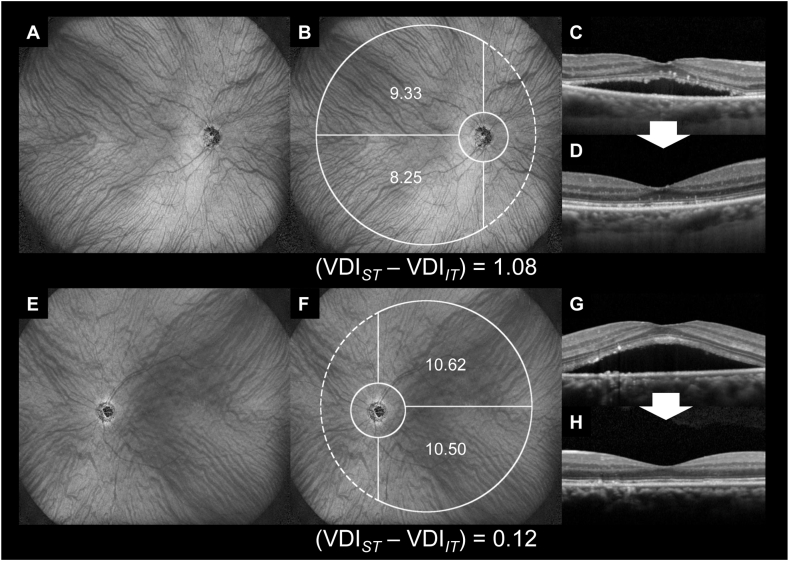
Twenty-nine patients with treatment-naïve CSC were divided into two groups based on the presence or absence of SRF 3 months after the initial visit (month 3) without any treatment. Using enhanced depth imaging of widefield swept-source optical coherence tomography, the choroidal thickness (CT), vessel density (VD), and vessel diameter index (VDI) in the superotemporal and inferotemporal subfields on the temporal side of the 18-mm circle from the disc were measured at the initial visit. We calculated the vertical difference in CT and other choroidal angiographic parameters and evaluated their association with the SRF condition at 3 months.

RESULTS

The SRF-resolved and SRF-persistent groups included 10 and 19 patients, respectively. At the initial visit, sex, age, axial length, symptom duration, the logarithm of the minimum angle of resolution visual acuity, and foveal thickness were not significantly different between the two groups. The SRF status at month 3 was not associated with the vertical difference in CT and choroidal VD ( = .614, .065, respectively). However, the vertical difference in choroidal VDI was positively associated with the future presence of SRF ( = .017).

CONCLUSIONS

Vertically asymmetric dilation of choroidal vessels in the posterior pole may be a vasculature feature associated with SRF from CSC and may be a good predictor of future SRF status.

摘要

目的

研究中心性浆液性脉络膜视网膜病变(CSC)患者后极部脉络膜血管造影特征与视网膜下液(SRF)消退或持续存在的关系。

设计

观察性病例系列。

方法

29例未经治疗的CSC患者在初次就诊后3个月(第3个月)未接受任何治疗,根据是否存在SRF分为两组。使用超广角扫频源光学相干断层扫描的增强深度成像,在初次就诊时测量距视盘18mm圆周颞侧的颞上和颞下子区域的脉络膜厚度(CT)、血管密度(VD)和血管直径指数(VDI)。我们计算了CT和其他脉络膜血管造影参数的垂直差异,并评估它们与3个月时SRF状况的相关性。

结果

SRF消退组和SRF持续组分别包括10例和19例患者。初次就诊时,两组在性别、年龄、眼轴长度、症状持续时间、最小分辨角视力的对数和黄斑厚度方面无显著差异。第3个月时的SRF状态与CT和脉络膜VD的垂直差异无关(分别为P = 0.614、0.065)。然而,脉络膜VDI的垂直差异与未来SRF的存在呈正相关(P = 0.017)。

结论

后极部脉络膜血管垂直不对称扩张可能是与CSC所致SRF相关的血管特征,可能是未来SRF状态的良好预测指标。

https://cdn.ncbi.nlm.nih.gov/pmc/blobs/b854/10412906/0c4d66910480/gr4.jpg
https://cdn.ncbi.nlm.nih.gov/pmc/blobs/b854/10412906/f3a98489e2f6/ga1.jpg
https://cdn.ncbi.nlm.nih.gov/pmc/blobs/b854/10412906/230620ba664b/gr1.jpg
https://cdn.ncbi.nlm.nih.gov/pmc/blobs/b854/10412906/a4a8bca239a3/gr2.jpg
https://cdn.ncbi.nlm.nih.gov/pmc/blobs/b854/10412906/92699e9374eb/gr3.jpg
https://cdn.ncbi.nlm.nih.gov/pmc/blobs/b854/10412906/0c4d66910480/gr4.jpg

相似文献

1
Widefield choroidal vasculature associated with future condition of subretinal fluid in central serous chorioretinopathy.中心性浆液性脉络膜视网膜病变中与未来视网膜下液状况相关的广角脉络膜血管系统。
Heliyon. 2023 Jul 22;9(8):e18441. doi: 10.1016/j.heliyon.2023.e18441. eCollection 2023 Aug.
2
Widefield Choroidal Thickness of Eyes with Central Serous Chorioretinopathy Examined by Swept-Source OCT.应用扫频源光学相干断层扫描仪测量中心性浆液性脉络膜视网膜病变眼的全视野脉络膜厚度。
Ophthalmol Retina. 2022 Oct;6(10):949-956. doi: 10.1016/j.oret.2022.04.011. Epub 2022 Apr 15.
3
Three-dimensional choroidal vascularity index in central serous chorioretinopathy using ultra-widefield swept-source optical coherence tomography angiography.使用超广角扫频源光学相干断层扫描血管造影术测量中心性浆液性脉络膜视网膜病变的三维脉络膜血管指数
Front Med (Lausanne). 2022 Sep 7;9:967369. doi: 10.3389/fmed.2022.967369. eCollection 2022.
4
Quantitative analysis of choroidal vasculature in central serous chorioretinopathy using ultra-widefield swept-source optical coherence tomography angiography.采用超广角扫频源光相干断层扫描血管造影术定量分析中心性浆液性脉络膜视网膜病变脉络膜血管。
Sci Rep. 2022 Nov 1;12(1):18427. doi: 10.1038/s41598-022-23389-1.
5
Optical Coherence Tomography Parameters as Predictors of Treatment Response to Eplerenone in Central Serous Chorioretinopathy.光学相干断层扫描参数作为中心性浆液性脉络膜视网膜病变中依普利酮治疗反应的预测指标
J Clin Med. 2019 Aug 22;8(9):1271. doi: 10.3390/jcm8091271.
6
Photodynamic therapy in chronic central serous chorioretinopathy with subretinal fluid outside the fovea.光动力疗法治疗黄斑中心凹外有视网膜下液的慢性中心性浆液性脉络膜视网膜病变。
Graefes Arch Clin Exp Ophthalmol. 2017 Oct;255(10):2029-2035. doi: 10.1007/s00417-017-3720-z. Epub 2017 Jul 3.
7
Abnormal choroidal flow on optical coherence tomography angiography in central serous chorioretinopathy.脉络膜血流异常在中心性浆液性脉络膜视网膜病变的光学相干断层扫描血管造影中的表现。
Clin Exp Ophthalmol. 2019 May;47(4):505-512. doi: 10.1111/ceo.13454. Epub 2019 Feb 17.
8
Association of Subretinal Fluid Duration and Baseline Chorioretinal Structure With Optical Coherence Tomography in Central Serous Chorioretinopathy.中心性浆液性脉络膜视网膜病变中视网膜下液持续时间和基线脉络膜视网膜结构与光相干断层扫描的相关性。
Transl Vis Sci Technol. 2023 Oct 3;12(10):12. doi: 10.1167/tvst.12.10.12.
9
Ultra-widefield imaging with autofluorescence and indocyanine green angiography in central serous chorioretinopathy.中心性浆液性脉络膜视网膜病变的超广角自体荧光和吲哚菁绿血管造影成像
Am J Ophthalmol. 2014 Aug;158(2):362-371.e2. doi: 10.1016/j.ajo.2014.04.021. Epub 2014 May 1.
10
Comparison of Retinal Layer Thickness and Capillary Vessel Density in the Patients with Spontaneously Resolved Acute Central Serous Chorioretinopathy.自发缓解的急性中心性浆液性脉络膜视网膜病变患者视网膜层厚度与毛细血管密度的比较
J Clin Med. 2020 Dec 25;10(1):45. doi: 10.3390/jcm10010045.

引用本文的文献

1
Six-month outcomes of half-fluence photodynamic therapy combined with intravitreal Aflibercept injection for pachychoroid neovasculopathy.半剂量光动力疗法联合玻璃体内注射阿柏西普治疗厚脉络膜新生血管病变的六个月疗效
Graefes Arch Clin Exp Ophthalmol. 2025 Aug 25. doi: 10.1007/s00417-025-06945-z.
2
Multimodal chorioretinal imaging in Wyburn-Mason syndrome: A case report.怀伯恩-梅森综合征的多模态脉络膜视网膜成像:一例报告
Heliyon. 2024 Jul 20;10(14):e34872. doi: 10.1016/j.heliyon.2024.e34872. eCollection 2024 Jul 30.
3
Sex differences in choroidal vessels using novel wide-field choroidal en-face images from optical coherence tomography.

本文引用的文献

1
Effectiveness of Reduced-fluence Photodynamic Therapy for Chronic Central Serous Chorioretinopathy: A Propensity Score Analysis.低能量光动力疗法治疗慢性中心性浆液性脉络膜视网膜病变的疗效:一项倾向评分分析
Ophthalmol Sci. 2022 Apr 6;2(2):100152. doi: 10.1016/j.xops.2022.100152. eCollection 2022 Jun.
2
A new insight into pachychoroid diseases: Remodeling of choroidal vasculature.对肥厚脉络膜疾病的新认识:脉络膜血管重构。
Graefes Arch Clin Exp Ophthalmol. 2022 Nov;260(11):3405-3417. doi: 10.1007/s00417-022-05687-6. Epub 2022 May 16.
3
Spontaneous Resolution of Chronic Central Serous Chorioretinopathy: "Fuji Sign".
利用光学相干断层扫描的新型广角脉络膜面图像观察脉络膜血管的性别差异。
Sci Rep. 2024 Jul 23;14(1):17013. doi: 10.1038/s41598-024-67671-w.
慢性中心性浆液性脉络膜视网膜病变的自发消退:“富士征”
Ophthalmol Retina. 2022 Sep;6(9):861-863. doi: 10.1016/j.oret.2022.04.023. Epub 2022 May 6.
4
Widefield Choroidal Thickness of Eyes with Central Serous Chorioretinopathy Examined by Swept-Source OCT.应用扫频源光学相干断层扫描仪测量中心性浆液性脉络膜视网膜病变眼的全视野脉络膜厚度。
Ophthalmol Retina. 2022 Oct;6(10):949-956. doi: 10.1016/j.oret.2022.04.011. Epub 2022 Apr 15.
5
CILIOCHOROIDAL EFFUSION IN CENTRAL SEROUS CHORIORETINOPATHY.脉络膜渗漏在中心性浆液性脉络膜视网膜病变中的表现。
Retina. 2022 Apr 1;42(4):730-737. doi: 10.1097/IAE.0000000000003376.
6
Clinical Factors Related to Loculation of Fluid in Central Serous Chorioretinopathy.与中心性浆液性脉络膜视网膜病变液体积聚相关的临床因素。
Am J Ophthalmol. 2022 Mar;235:197-203. doi: 10.1016/j.ajo.2021.09.009. Epub 2021 Sep 20.
7
Incidence of central serous chorioretinopathy (2011-2018): a nationwide population-based cohort study of Japan.中心性浆液性脉络膜视网膜病变的发病率(2011-2018 年):日本一项基于全国人口的队列研究。
Br J Ophthalmol. 2022 Dec;106(12):1748-1753. doi: 10.1136/bjophthalmol-2021-319403. Epub 2021 Jul 14.
8
Venous overload choroidopathy: A hypothetical framework for central serous chorioretinopathy and allied disorders.静脉超负荷性脉络膜病变:中心性浆液性脉络膜视网膜病变及相关疾病的一个假说框架
Prog Retin Eye Res. 2022 Jan;86:100973. doi: 10.1016/j.preteyeres.2021.100973. Epub 2021 May 21.
9
ANGIOGRAPHIC RISK FACTORS FOR RECURRENCE OF MACULAR EDEMA ASSOCIATED WITH BRANCH RETINAL VEIN OCCLUSION.与分支视网膜静脉阻塞相关的黄斑水肿复发的血管造影危险因素。
Retina. 2021 Jun 1;41(6):1219-1226. doi: 10.1097/IAE.0000000000003026.
10
Vortex Vein Anastomosis at the Watershed in Pachychoroid Spectrum Diseases.脉络膜增厚谱病变中涡静脉吻合在分水岭区。
Ophthalmol Retina. 2020 Sep;4(9):938-945. doi: 10.1016/j.oret.2020.03.024. Epub 2020 Apr 6.