Osaku Daiken, Taniguchi Fuminori, Komatsu Hiroaki, Wibisono Hermawan, Azuma Yukihiro, Harada Tasuku
Department of Obstetrics and Gynecology, Tottori University Faculty of Medicine, Yonago, Japan.
Gynecol Minim Invasive Ther. 2021 Nov 5;10(4):256-258. doi: 10.4103/GMIT.GMIT_16_20. eCollection 2021 Oct-Dec.
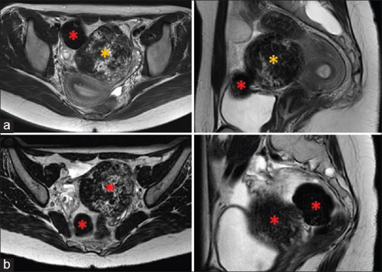
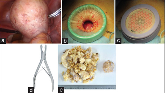
Basal cell nevus syndrome (BCNS) is a rare neurocutaneous syndrome characterized by tumorigeneses such as basal cell carcinomas, jaw cysts, ovarian fibromas, and cardiac fibromas. We present a 24-year-old female with calcified ovarian fibromas associated with BCNS. She had a surgical history of the maxillary cyst and was diagnosed with BCNS due to the cutaneous pits. Magnetic resonance imaging indicated an 8-cm mass and a 4-cm mass, which had been suspected to be a subserosal myoma and a fibroma, respectively. GnRH agonist was preoperatively administered; however, the size of the masses did not change. In laparoscopy, a tumor consisting of 8- and 5-cm masses in the right ovary was identified, and tumorectomy was performed. Because both tumors were extraordinarily rigid and could not be morcellated with scalpels or scissors, we removed them by the Luer Bone Rongeurs with minilaparotomy. The histopathological diagnosis was the ovarian fibromas with marked calcification.
基底细胞痣综合征(BCNS)是一种罕见的神经皮肤综合征,其特征为发生诸如基底细胞癌、颌骨囊肿、卵巢纤维瘤和心脏纤维瘤等肿瘤。我们报告一例24岁患有与BCNS相关的钙化性卵巢纤维瘤的女性。她有上颌囊肿手术史,因皮肤凹陷被诊断为BCNS。磁共振成像显示一个8厘米肿物和一个4厘米肿物,分别怀疑为浆膜下肌瘤和纤维瘤。术前给予促性腺激素释放激素(GnRH)激动剂;然而,肿物大小未改变。在腹腔镜检查中,发现右侧卵巢有一个由8厘米和5厘米肿物组成的肿瘤,并进行了肿瘤切除术。由于两个肿瘤都异常坚硬,无法用手术刀或剪刀切碎,我们通过小切口剖腹术用鲁尔咬骨钳将它们取出。组织病理学诊断为伴有明显钙化的卵巢纤维瘤。