Kobayashi Hisashi, Imai Yasuo, Hirao Takayuki, Nakao Ko, Kajinaka Hayato, Kishi Kazuo
Department of Plastic and Reconstructive Surgery, Teikyo University Chiba Medical Center, Chiba, Japan.
Department of Diagnostic Pathology, Ota Memorial Hospital, Gunma, Japan.
Plast Reconstr Surg Glob Open. 2021 Dec 20;9(12):e3967. doi: 10.1097/GOX.0000000000003967. eCollection 2021 Dec.
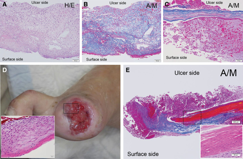
Decellularized porcine small intestinal submucosa (SIS), commercialized as an extracellular matrix rich in cell-inducing substrates and factors, has been clinically applied to treat intractable skin ulcers and has shown therapeutic effects. The SIS reportedly induces cell infiltration and integrates with the ulcer bed after 3-7 days of application. The attached SIS degenerates over time, and the remaining mass appears as slough, below which is granulation tissue that is essential for healing. This study aimed to determine whether the slough should be removed in clinical settings.
Five patients with intractable skin ulcers were included in this case series. Seven days after applying a two-layer fenestrated-type SIS to the ulcer, the removed slough was histopathologically examined.
The collagen fibers of the SIS somewhat degenerated, and inflammatory cell infiltration was observed from the ulcer side to the surface side of the SIS. Neovascularization was similarly observed on the ulcer side. The degree of inflammatory cell infiltration decreased from the ulcer side to the surface side, whereas pus (ie, aggregates of neutrophils) was observed on the surface and ulcer edges. Additionally, the removed slough contained regenerative epithelium on the ulcer side of the remaining collagen fibers.
After treating intractable skin ulcers using SIS, we recommend removal of the upper surface and ulcer edge of the degenerated SIS or slough to prevent infection and preservation of the lower side of the degenerated SIS to maintain the granulation tissue and regenerative epithelium.
脱细胞猪小肠黏膜下层(SIS)作为一种富含细胞诱导底物和因子的细胞外基质已商业化,已临床应用于治疗顽固性皮肤溃疡并显示出治疗效果。据报道,SIS在应用3 - 7天后可诱导细胞浸润并与溃疡床整合。附着的SIS会随着时间退化,剩余物质表现为腐痂,其下方是愈合所必需的肉芽组织。本研究旨在确定在临床环境中是否应去除腐痂。
本病例系列纳入了5例顽固性皮肤溃疡患者。在将两层开窗型SIS应用于溃疡7天后,对切除的腐痂进行组织病理学检查。
SIS的胶原纤维有所退化,从溃疡侧向SIS表面侧观察到炎性细胞浸润。在溃疡侧同样观察到新生血管形成。炎性细胞浸润程度从溃疡侧向表面侧降低,而在表面和溃疡边缘观察到脓液(即中性粒细胞聚集物)。此外,切除的腐痂在剩余胶原纤维的溃疡侧含有再生上皮。
使用SIS治疗顽固性皮肤溃疡后,我们建议去除退化的SIS或腐痂的上表面和溃疡边缘以预防感染,并保留退化SIS的下侧以维持肉芽组织和再生上皮。