Malviya Kapil Kumar, Verma Ashish, Nayak Amit Kumar, Mishra Anand, More Raghunath Shahaji
Department of Anatomy, Institute of Medical Science, Banaras Hindu University, Varanasi 221005, India.
Department of Radiodiagnosis and Imaging, Institute of Medical Science, Banaras Hindu University, Varanasi 221005, India.
Diagnostics (Basel). 2021 Dec 3;11(12):2262. doi: 10.3390/diagnostics11122262.
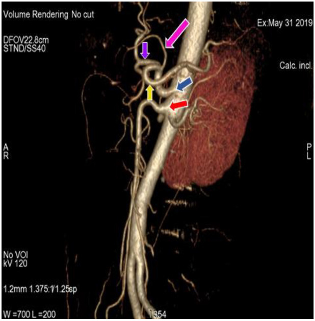
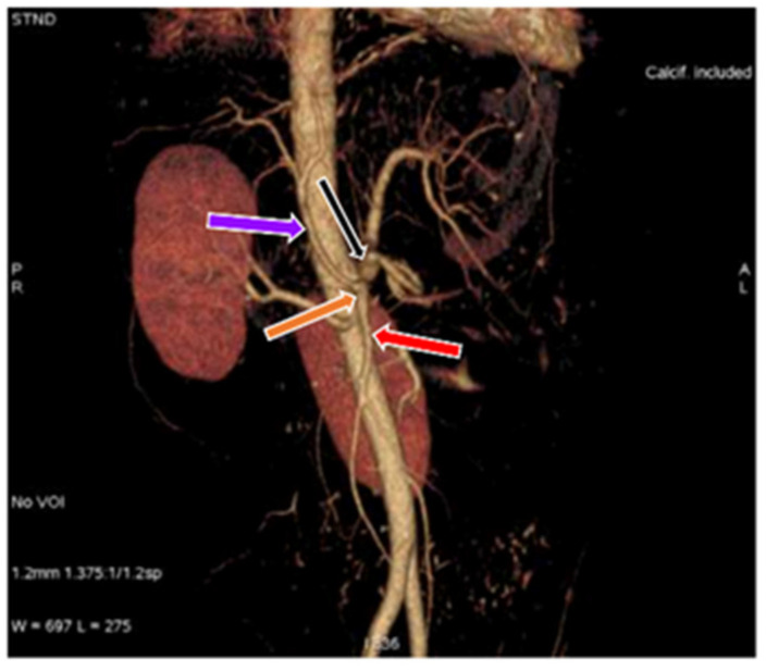
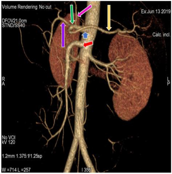
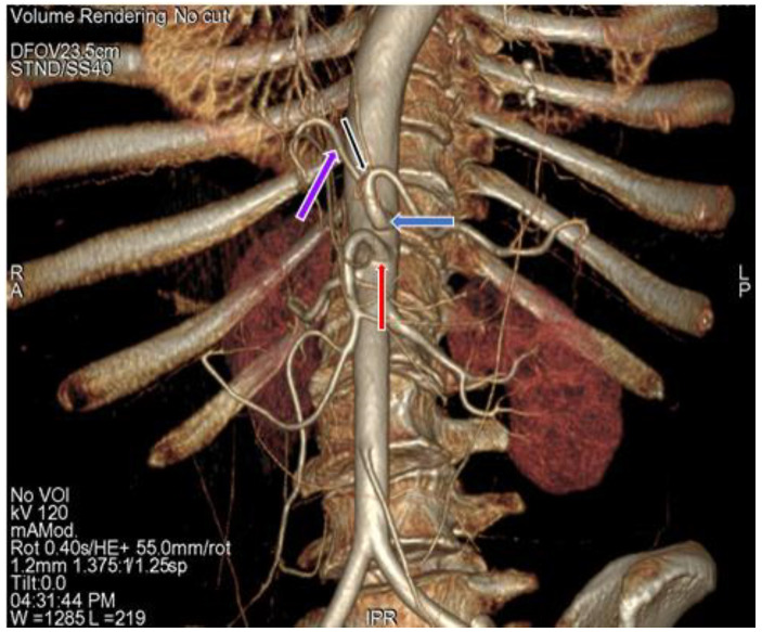
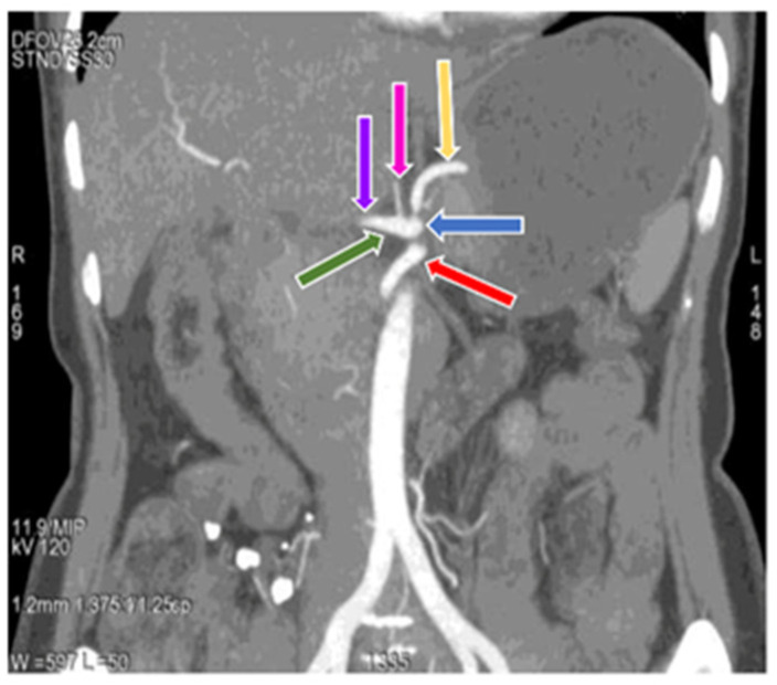
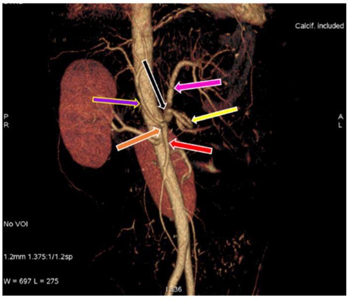
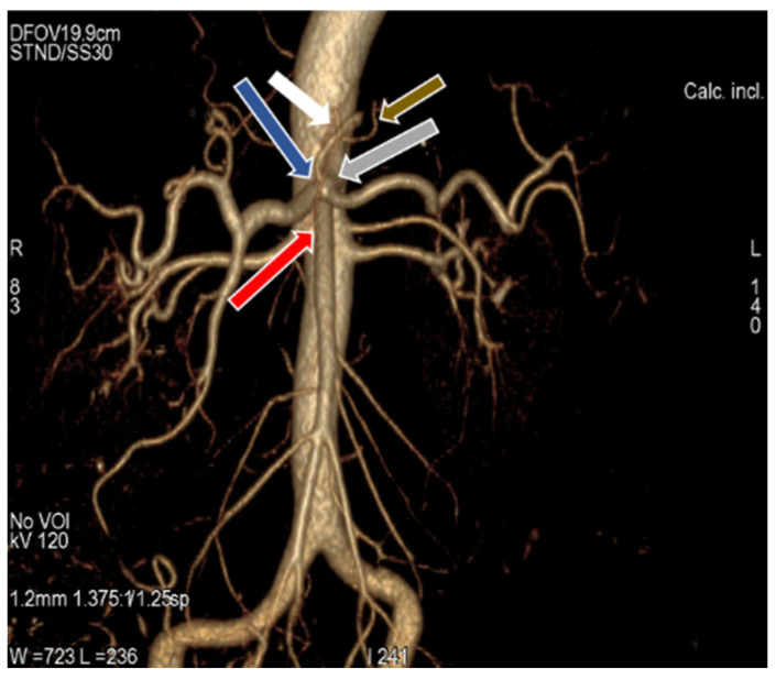
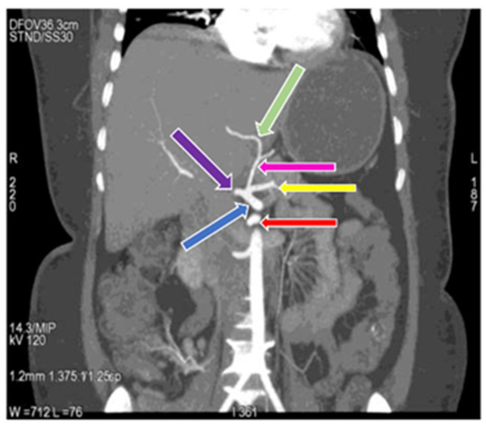
Understanding of variations in the course and source of abdominal arteries is crucial for any surgical intervention in the peritoneal space. Intricate surgeries of the upper abdominal region, such as hepato-biliary, pancreatic, gastric and splenic surgeries, require precise knowledge of regular anatomy and different variations related to celiac trunk and hepatic artery. In addition, information about the origin of inferior phrenic artery is important in conditions such as hepatocellular carcinoma and gastroesophageal bleeding management. The present study gives an account of anatomical variations in origin and branching pattern of celiac trunk and hepatic artery by the use of CT (computed tomographic) angiography. The study was performed on 110 (66 females and 44 males) patients in a north Indian population. Results unraveled the most common celiac trunk variation as hepatosplenic trunk with left gastric artery, which was observed in 60% of cases, more common in females than in males. Gastrosplenic and hepato-gastric trunk could be seen in 4.55% and 1.82% cases respectively. Gastrosplenic trunk was more commonly found in females, whereas hepato-gastric trunk was more common in males. A gastrosplenic trunk, along with the hepato-mesenteric trunk, was observed in 1.82% cases and was more common in males. A celiacomesenteric trunk, in which the celiac trunk and superior mesenteric artery originated as a common trunk from the aorta, was seen only in 0.91% of cases, and exhibited an origin of right and left inferior phrenic artery from the left gastric artery. The most common variation of hepatic artery, in which the right hepatic artery was replaced and originated from the superior mesenteric artery, was observed in 3.64%, cases with a more common occurrence in males. In 1.82% cases, the left hepatic artery was replaced and originated from the left gastric artery, which was observed only in females. Common hepatic artery originated from the superior mesenteric artery, as observed in 1.82% cases, with slightly higher occurrence in males. These findings not only add to the existing knowledge apart from giving an overview of variations in north Indian population, but also give an account of their correlation with gender. The present study will prove to be important for various surgeries of the upper abdominal region.
了解腹主动脉的走行和来源的变异对于腹膜腔的任何手术干预都至关重要。上腹部区域的复杂手术,如肝胆、胰腺、胃和脾脏手术,需要精确掌握与腹腔干和肝动脉相关的正常解剖结构及不同变异。此外,膈下动脉起源的信息在肝细胞癌和胃食管出血管理等病症中很重要。本研究通过使用CT(计算机断层扫描)血管造影描述了腹腔干和肝动脉起源及分支模式的解剖变异。该研究在印度北部人群的110名患者(66名女性和44名男性)中进行。结果揭示最常见的腹腔干变异是肝脾干与胃左动脉,在60%的病例中观察到,女性比男性更常见。胃脾干和肝胃干分别在4.55%和1.82%的病例中可见。胃脾干在女性中更常见,而肝胃干在男性中更常见。在1.82%的病例中观察到胃脾干与肝肠系膜干,在男性中更常见。腹腔肠系膜干,即腹腔干和肠系膜上动脉从主动脉发出形成一个共同干,仅在0.91%的病例中可见,并表现出左右膈下动脉从胃左动脉发出。最常见的肝动脉变异,即右肝动脉被替代并起源于肠系膜上动脉,在3.64%的病例中观察到,男性中更常见。在1.82%的病例中,左肝动脉被替代并起源于胃左动脉,仅在女性中观察到。肝总动脉起源于肠系膜上动脉,在1.82%的病例中观察到,男性中发生率略高。这些发现不仅除了概述印度北部人群的变异外还增加了现有知识,而且还说明了它们与性别的相关性。本研究将对上腹部区域的各种手术具有重要意义。