Botermans Anna, Lidén Anna, de Carvalho Machado Vinícius, Chrcanovic Bruno Ramos
Faculty of Odontology, Malmö University, 214 21 Malmö, Sweden.
Slice Diagnóstico Volumétrico por Imagem, Belo Horizonte 30140-110, Brazil.
J Clin Med. 2021 Dec 14;10(24):5853. doi: 10.3390/jcm10245853.
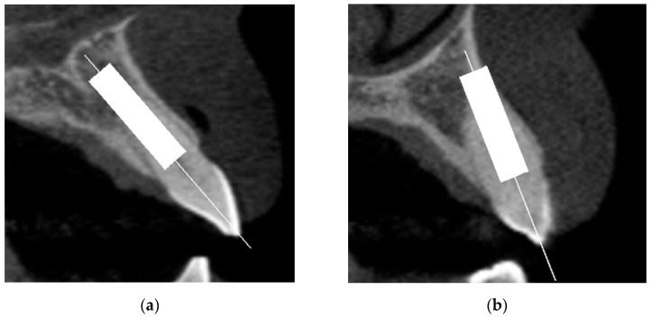
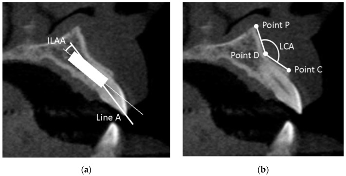
This study aimed to investigate the factors that could be associated with the risk of labial cortical bone wall perforation with immediate implant placement (IIP) in the maxillary aesthetic zone, in a cone-beam computed tomography (CBCT) virtual study. CBCT exams from 126 qualified subjects (756 teeth) were included. Implants were virtually positioned in two different positions: in the long axis of the tooth (prosthetically-driven position) and in an ideal position in relation to adjacent anatomical structures (bone-driven position). Two different implant diameters were planned for each tooth position, namely, 3.75 and 4.3 mm for central incisors and canines, and 3.0 and 3.3 mm for lateral incisors. The incidence of perforation was nearly 80% and 5% for prosthetically- and bone-driven position, respectively. Factors associated with a higher risk of cortical bone wall perforation (bone-driven position), according to logistic regression analysis, were women, wider implants, Sagittal Root Position class IV, and decrease of the labial concavity angle. Perforation of the labial cortical bone wall can be greatly minimized when the implant is placed in a bone-driven position compared to a prosthetically-driven position. It is important to preoperatively evaluate the morphological features of the implant site for risk assessment and to individualize the treatment plan.
本研究旨在通过锥束计算机断层扫描(CBCT)虚拟研究,调查在上颌美学区即刻种植(IIP)时可能与唇侧皮质骨壁穿孔风险相关的因素。纳入了126名合格受试者(756颗牙齿)的CBCT检查。种植体虚拟定位在两个不同位置:沿牙齿长轴(修复驱动位置)和相对于相邻解剖结构的理想位置(骨驱动位置)。为每个牙齿位置规划了两种不同的种植体直径,即中切牙和尖牙为3.75和4.3毫米,侧切牙为3.0和3.3毫米。修复驱动位置和骨驱动位置的穿孔发生率分别约为80%和5%。根据逻辑回归分析,与皮质骨壁穿孔风险较高(骨驱动位置)相关的因素为女性、较宽的种植体、矢状根位置IV类以及唇侧凹陷角度减小。与修复驱动位置相比,当种植体置于骨驱动位置时,唇侧皮质骨壁穿孔可大大减少。术前评估种植部位的形态特征以进行风险评估并制定个体化治疗方案很重要。