Department of Advanced General Dentistry, Faculty of Dentistry, Mahidol University, Yothi Road, Ratchathewi District, Bangkok 10400, Thailand.
Department of Oral and Maxillofacial Radiology, Faculty of Dentistry, Mahidol University, Bangkok 10400, Thailand.
Molecules. 2022 Jan 18;27(3):608. doi: 10.3390/molecules27030608.
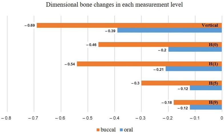
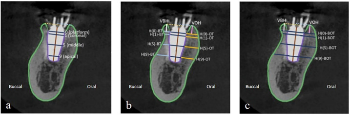
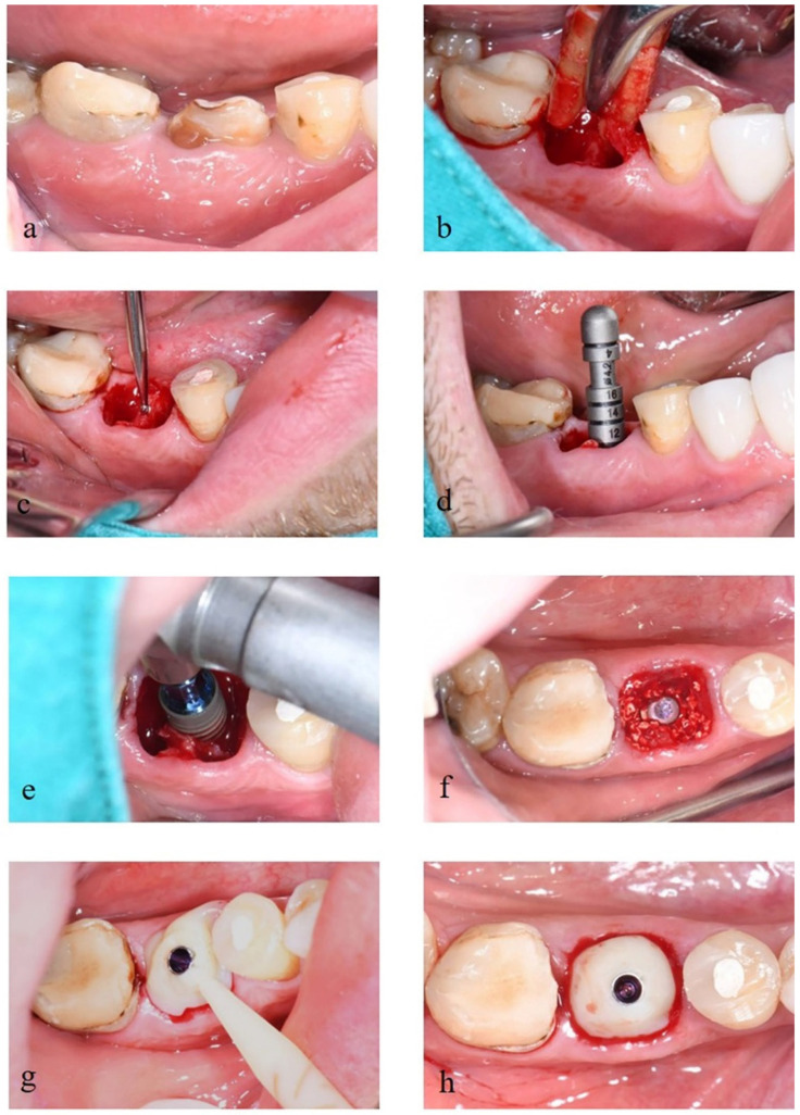
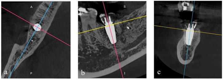
This prospective clinical study aimed to evaluate the peri-implant hard tissue dimensional change at 6 months of immediate implant placement with bone graft materials in the posterior area using cone-beam computed tomography (CBCT). Twelve dental implants were placed concurrently following tooth extraction in the posterior area and filled with xenograft particles. The CBCT images were taken immediately after surgical procedures and then at 6 months follow-up. To evaluate the hard tissue changes, the vertical and horizontal bone thickness were analyzed and measured using ImageJ software. Paired -test or Wilcoxon match-pair signed-rank test was done to analyze the changes of hard tissue values at the same level between immediately and 6 months following immediate implant placement. Independent -test or Mann-Whitney U test was used to analyze the dimensional change in the vertical and horizontal direction in buccal and lingual aspects. The level of significance was set at value = 0.05. All implants were successfully osseointegrated. At 6 months follow-up, the vertical bone change at the buccal aspect was -0.69 mm and at the lingual aspect -0.39 mm. For horizontal bone thickness, the bone dimensional changes at 0, 1, 5, and 9 mm levels from the implant platform were -0.62 mm, -0.70 mm, -0.24 mm, and -0.22 mm, respectively. A significant bone reduction was observed in all measurement levels during the 6 months after implant placement ( value < 0.05). It was noted that even with bone grafting, a decrease in bone thickness was seen following the immediate implant placement. Therefore, this technique can be an alternative method to place the implant in the posterior area.
本前瞻性临床研究旨在使用锥形束计算机断层扫描 (CBCT) 评估在后牙区使用骨移植材料即刻植入时的种植体周围硬组织在 6 个月时的尺寸变化。12 颗牙种植体在拔牙后同期植入,并填充异种移植物颗粒。在手术完成后立即和 6 个月随访时拍摄 CBCT 图像。使用 ImageJ 软件分析和测量硬组织变化,以评估垂直和水平骨厚度。使用配对检验或 Wilcoxon 匹配对符号秩检验分析即刻植入和即刻植入后 6 个月同一水平的硬组织值变化。使用独立检验或 Mann-Whitney U 检验分析颊舌方向的垂直和水平方向的尺寸变化。显著性水平设置为 值= 0.05。所有种植体均成功骨整合。在 6 个月随访时,颊侧的垂直骨变化为-0.69mm,舌侧为-0.39mm。对于水平骨厚度,从种植体平台的 0、1、5 和 9mm 水平的骨尺寸变化分别为-0.62mm、-0.70mm、-0.24mm 和-0.22mm。在种植体放置后的 6 个月内,所有测量水平均观察到明显的骨质减少( 值<0.05)。值得注意的是,即使进行了骨移植,在即刻植入后也会看到骨厚度减少。因此,这种技术可以成为在后牙区放置种植体的替代方法。