Lou Jieqiong, Solano Ashleigh, Liang Zhen, Hinde Elizabeth
School of Physics, University of Melbourne, Melbourne, VIC, Australia.
Department of Biochemistry and Pharmacology, University of Melbourne, Melbourne, VIC, Australia.
Front Genet. 2021 Dec 10;12:770081. doi: 10.3389/fgene.2021.770081. eCollection 2021.
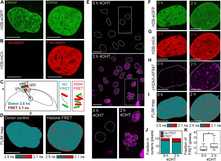
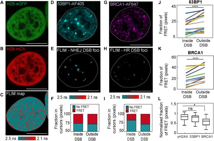
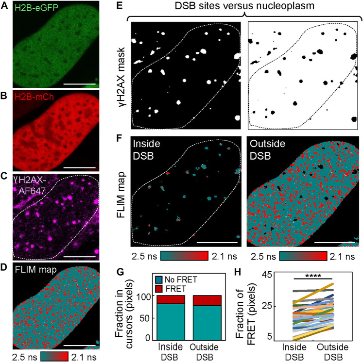
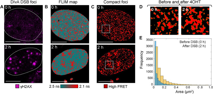
A DNA double-strand break (DSB) takes place in the context of chromatin, and there is increasing evidence for chromatin structure to play a functional role in DSB signaling and repair. Thus, there is an emerging need for quantitative microscopy methods that can directly measure chromatin network architecture and detect changes in this structural framework upon DSB induction within an intact nucleus. To address this demand, here we present the phasor approach to fluorescence lifetime imaging microscopy (FLIM) of Förster resonance energy transfer (FRET) between fluorescently labeled histones in the DSB inducible via AsiSI cell system (DIvA), which has sufficient spatial resolution to map nuclear-wide chromatin compaction at the level of nucleosome proximity with respect to multiple site-specific DSBs. We also demonstrate that when phasor histone FLIM-FRET is coupled with immunofluorescence, this technology has the unique advantage of enabling exploration of any heterogeneity that exists in chromatin structure at the spatially distinct and genetically induced DSBs.
DNA双链断裂(DSB)发生在染色质环境中,越来越多的证据表明染色质结构在DSB信号传导和修复中发挥功能作用。因此,迫切需要定量显微镜方法,能够直接测量染色质网络结构,并检测完整细胞核内DSB诱导后这种结构框架的变化。为满足这一需求,我们在此介绍通过AsiSI细胞系统(DIvA)诱导DSB时,荧光标记组蛋白之间Förster共振能量转移(FRET)的荧光寿命成像显微镜(FLIM)的相量方法,该方法具有足够的空间分辨率,能够在核小体接近水平上绘制全核染色质压实情况,涉及多个位点特异性DSB。我们还证明,当相量组蛋白FLIM-FRET与免疫荧光相结合时,这项技术具有独特优势,能够探索在空间上不同且由基因诱导的DSB处染色质结构中存在的任何异质性。