Department of Reproduction, Obstetrics and Herd Health, Faculty of Veterinary Medicine, Ghent University, Ghent, Belgium.
Department of Clinical Sciences, Utrecht University, Utrecht, The Netherlands.
Biol Reprod. 2022 Apr 26;106(4):710-729. doi: 10.1093/biolre/ioab243.
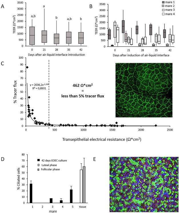
We describe the development of two methods for obtaining confluent monolayers of polarized, differentiated equine oviduct epithelial cells (EOEC) in Transwell inserts and microfluidic chips. EOECs from the ampulla were isolated post-mortem and seeded either (1) directly onto a microporous membrane as differentiated EOECs (direct seeding protocol) or (2) first cultured to a confluent de-differentiated monolayer in conventional wells, then trypsinized and seeded onto a microporous membrane (re-differentiation protocol). Maintenance or induction of EOEC differentiation in these systems was achieved by air-liquid interface introduction. Monolayers cultured via both protocols were characterized by columnar, cytokeratin 19-positive EOECs in Transwell inserts. However, only the re-differentiation protocol could be transferred successfully to the microfluidic chips. Integrity of the monolayers was confirmed by transepithelial resistance measurements, tracer flux, and the demonstration of an intimate network of tight junctions. Using the direct protocol, 28% of EOECs showed secondary cilia at the apical surface in a diffuse pattern. In contrast, re-differentiated polarized EOECs rarely showed secondary cilia in either culture system (>90% of the monolayers showed <1% ciliated EOECs). Occasionally (5-10%), re-differentiated monolayers with 11-27% EOECs with secondary cilia in a diffuse pattern were obtained. Additionally, nuclear progesterone receptor expression was found to be inhibited by simulated luteal phase hormone concentrations, and sperm binding to cilia was higher for re-differentiated EOEC monolayers exposed to estrogen-progesterone concentrations mimicking the follicular rather than luteal phase. Overall, a functional equine oviduct model was established with close morphological resemblance to in vivo oviduct epithelium.
我们描述了两种方法的发展,用于获得在 Transwell 插入物和微流控芯片中具有极化和分化的马输卵管上皮细胞 (EOEC) 的融合单层。死后从壶腹分离 EOEC 并直接接种到微孔膜上(分化 EOEC 的直接接种方案),或者(2)首先在常规培养皿中培养至融合的去分化单层,然后用胰蛋白酶消化并接种到微孔膜上(重新分化方案)。在这些系统中通过气液界面引入来维持或诱导 EOEC 分化。通过这两种方案培养的单层在 Transwell 插入物中表现为柱状、细胞角蛋白 19 阳性的 EOEC。然而,只有重新分化方案才能成功转移到微流控芯片上。通过跨上皮电阻测量、示踪剂通量和紧密连接的紧密网络的证明来确认单层的完整性。使用直接方案,28%的 EOEC 在弥漫模式的顶表面显示出次级纤毛。相比之下,重新分化的极化 EOEC 在这两种培养系统中很少显示次级纤毛(>90%的单层显示<1%有纤毛的 EOEC)。偶尔(5-10%),在重新分化的单层中获得 11-27%具有弥漫模式次级纤毛的 EOEC。此外,发现模拟黄体期激素浓度抑制核孕激素受体表达,并且对于暴露于模拟卵泡期而不是黄体期的雌激素孕激素浓度的重新分化的 EOEC 单层,精子与纤毛的结合更高。总体而言,建立了具有与体内输卵管上皮非常相似的形态学特征的功能性马输卵管模型。