• 文献检索
  • 文档翻译
  • 深度研究
  • 学术资讯
  • Suppr Zotero 插件Zotero 插件
  • 邀请有礼
  • 套餐&价格
  • 历史记录
应用&插件
Suppr Zotero 插件Zotero 插件浏览器插件Mac 客户端Windows 客户端微信小程序
定价
高级版会员购买积分包购买API积分包
服务
文献检索文档翻译深度研究API 文档MCP 服务
关于我们
关于 Suppr公司介绍联系我们用户协议隐私条款
关注我们

Suppr 超能文献

核心技术专利:CN118964589B侵权必究
粤ICP备2023148730 号-1Suppr @ 2026

文献检索

告别复杂PubMed语法,用中文像聊天一样搜索,搜遍4000万医学文献。AI智能推荐,让科研检索更轻松。

立即免费搜索

文件翻译

保留排版,准确专业,支持PDF/Word/PPT等文件格式,支持 12+语言互译。

免费翻译文档

深度研究

AI帮你快速写综述,25分钟生成高质量综述,智能提取关键信息,辅助科研写作。

立即免费体验

甲状旁腺激素信号在成熟成骨细胞/骨细胞中保护小鼠免受与年龄相关的骨丢失。

Parathyroid hormone signaling in mature osteoblasts/osteocytes protects mice from age-related bone loss.

机构信息

Department of Translational Dental Medicine, Goldman School of Dental Medicine, Boston University, Boston, MA 02118, USA.

Endocrine Unit, Massachusetts General Hospital, Harvard Medical School, Boston, MA 02114, USA.

出版信息

Aging (Albany NY). 2021 Dec 30;13(24):25607-25642. doi: 10.18632/aging.203808.

DOI:10.18632/aging.203808
PMID:34968192
原文链接:https://pmc.ncbi.nlm.nih.gov/articles/PMC8751595/
Abstract

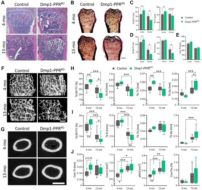
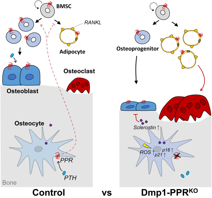
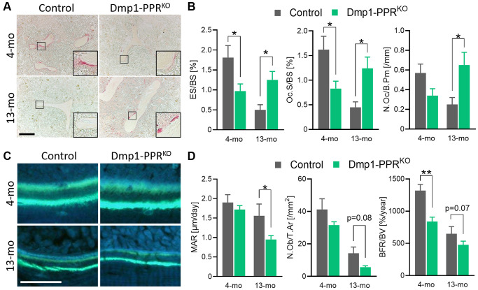
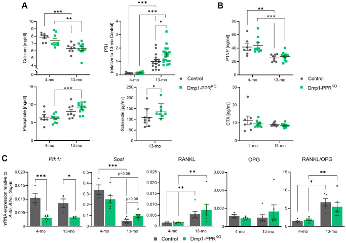
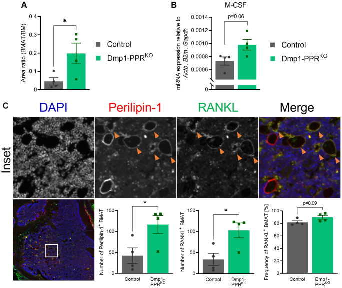
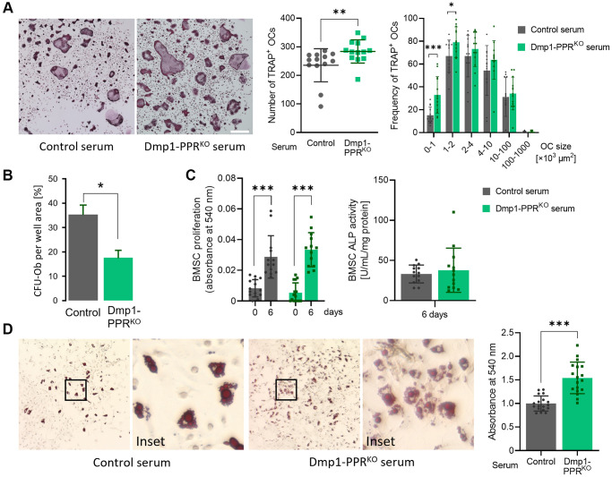
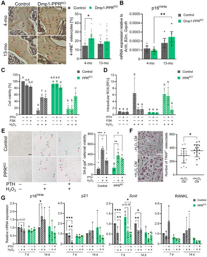
Aging is accompanied by osteopenia, characterized by reduced bone formation and increased bone resorption. Osteocytes, the terminally differentiated osteoblasts, are regulators of bone homeostasis, and parathyroid hormone (PTH) receptor (PPR) signaling in mature osteoblasts/osteocytes is essential for PTH-driven anabolic and catabolic skeletal responses. However, the role of PPR signaling in those cells during aging has not been investigated. The aim of this study was to analyze the role of PTH signaling in mature osteoblasts/osteocytes during aging. Mice lacking PPR in osteocyte (Dmp1-PPR) display an age-dependent osteopenia characterized by a significant decrease in osteoblast activity and increase in osteoclast number and activity. At the molecular level, the absence of PPR signaling in mature osteoblasts/osteocytes is associated with an increase in serum sclerostin and a significant increase in osteocytes expressing 4-hydroxy-2-nonenals, a marker of oxidative stress. In Dmp1-PPR mice there was an age-dependent increase in p16/ expression, whereas it was unchanged in controls. studies demonstrated that PTH protects osteocytes from oxidative stress-induced cell death. In summary, we reported that PPR signaling in osteocytes is important for protecting the skeleton from age-induced bone loss by restraining osteoclast's activity and protecting osteocytes from oxidative stresses.

摘要

衰老是伴随着骨质疏松症的,其特征是骨形成减少和骨吸收增加。成骨细胞是终末分化的成骨细胞,是骨稳态的调节者,成熟成骨细胞/成骨细胞中的甲状旁腺激素 (PTH) 受体 (PPR) 信号对于 PTH 驱动的合成代谢和分解代谢骨骼反应是必不可少的。然而,PPR 信号在衰老过程中成骨细胞中的作用尚未被研究。本研究旨在分析 PTH 信号在成熟成骨细胞/成骨细胞中的作用衰老。缺乏成骨细胞中 PPR 的小鼠(Dmp1-PPR)表现出与年龄相关的骨质疏松症,其特征是成骨细胞活性显著降低,破骨细胞数量和活性增加。在分子水平上,成熟成骨细胞/成骨细胞中 PPR 信号的缺失与血清骨硬化素的增加以及表达 4-羟基-2-壬烯醛的成骨细胞数量的显著增加有关,这是氧化应激的标志物。在 Dmp1-PPR 小鼠中,p16/的表达随年龄增长而增加,而在对照组中则保持不变。研究表明,PTH 可保护成骨细胞免受氧化应激诱导的细胞死亡。总之,我们报告称,成骨细胞中的 PPR 信号对于通过抑制破骨细胞的活性和保护成骨细胞免受氧化应激来保护骨骼免受年龄相关的骨质流失是重要的。

https://cdn.ncbi.nlm.nih.gov/pmc/blobs/4b58/8751595/a588ae9c8088/aging-13-203808-g007.jpg
https://cdn.ncbi.nlm.nih.gov/pmc/blobs/4b58/8751595/df17ad06fefa/aging-13-203808-g001.jpg
https://cdn.ncbi.nlm.nih.gov/pmc/blobs/4b58/8751595/f6787372f847/aging-13-203808-g002.jpg
https://cdn.ncbi.nlm.nih.gov/pmc/blobs/4b58/8751595/ab0f974f7aa4/aging-13-203808-g003.jpg
https://cdn.ncbi.nlm.nih.gov/pmc/blobs/4b58/8751595/8c94375620f3/aging-13-203808-g004.jpg
https://cdn.ncbi.nlm.nih.gov/pmc/blobs/4b58/8751595/f8b4bafcc671/aging-13-203808-g005.jpg
https://cdn.ncbi.nlm.nih.gov/pmc/blobs/4b58/8751595/44c55020eef3/aging-13-203808-g006.jpg
https://cdn.ncbi.nlm.nih.gov/pmc/blobs/4b58/8751595/a588ae9c8088/aging-13-203808-g007.jpg

相似文献

1
Parathyroid hormone signaling in mature osteoblasts/osteocytes protects mice from age-related bone loss.甲状旁腺激素信号在成熟成骨细胞/骨细胞中保护小鼠免受与年龄相关的骨丢失。
Aging (Albany NY). 2021 Dec 30;13(24):25607-25642. doi: 10.18632/aging.203808.
2
Parathyroid hormone (PTH)/PTH-related peptide type 1 receptor (PPR) signaling in osteocytes regulates anabolic and catabolic skeletal responses to PTH.甲状旁腺激素(PTH)/甲状旁腺激素相关肽 1 型受体(PPR)在骨细胞中的信号转导调节 PTH 对骨骼的合成代谢和分解代谢反应。
J Biol Chem. 2013 Jul 12;288(28):20122-34. doi: 10.1074/jbc.M112.441360. Epub 2013 Jun 2.
3
Disruption of PTH receptor 1 in T cells protects against PTH-induced bone loss.破骨细胞 PTH 受体 1 可预防 PTH 诱导的骨丢失。
PLoS One. 2010 Aug 20;5(8):e12290. doi: 10.1371/journal.pone.0012290.
4
Parathyroid Hormone Induces Bone Cell Motility and Loss of Mature Osteocyte Phenotype through L-Calcium Channel Dependent and Independent Mechanisms.甲状旁腺激素通过L-钙通道依赖性和非依赖性机制诱导骨细胞运动并导致成熟骨细胞表型丧失。
PLoS One. 2015 May 5;10(5):e0125731. doi: 10.1371/journal.pone.0125731. eCollection 2015.
5
Resorption controls bone anabolism driven by parathyroid hormone (PTH) receptor signaling in osteocytes.破骨细胞吸收活动通过甲状旁腺激素(PTH)受体信号来控制成骨细胞中的骨合成代谢。
J Biol Chem. 2013 Oct 11;288(41):29809-20. doi: 10.1074/jbc.M113.485938. Epub 2013 Aug 20.
6
Loss of Gsα in osteocytes leads to osteopenia due to sclerostin induced suppression of osteoblast activity.成骨细胞中 Gsα 的缺失会导致骨量减少,这是由于骨钙素诱导的成骨细胞活性抑制所致。
Bone. 2018 Dec;117:138-148. doi: 10.1016/j.bone.2018.09.021. Epub 2018 Sep 25.
7
Control of Bone Anabolism in Response to Mechanical Loading and PTH by Distinct Mechanisms Downstream of the PTH Receptor.甲状旁腺激素(PTH)受体下游不同机制对机械负荷和PTH应答时骨合成代谢的调控
J Bone Miner Res. 2017 Mar;32(3):522-535. doi: 10.1002/jbmr.3011. Epub 2016 Oct 27.
8
PTH receptor signaling in osteocytes governs periosteal bone formation and intracortical remodeling.成骨细胞中甲状旁腺激素受体信号转导调控骨膜骨形成和皮质内骨重塑。
J Bone Miner Res. 2011 May;26(5):1035-46. doi: 10.1002/jbmr.304.
9
MMP14 is a novel target of PTH signaling in osteocytes that controls resorption by regulating soluble RANKL production.MMP14 是破骨细胞中 PTH 信号的一个新靶点,通过调节可溶性 RANKL 的产生来控制吸收。
FASEB J. 2018 May;32(5):2878-2890. doi: 10.1096/fj.201700919RRR. Epub 2018 Jan 17.
10
Low-dose PTH increases osteoblast activity via decreased Mef2c/Sost in senescent osteopenic mice.低剂量甲状旁腺激素通过降低衰老性骨质疏松小鼠的Mef2c/Sost来增加成骨细胞活性。
J Endocrinol. 2014 Oct;223(1):25-33. doi: 10.1530/JOE-14-0249. Epub 2014 Jul 23.

引用本文的文献

1
Caveolin-1 Regulates Parathyroid Hormone (PTH)-Related Protein (PTHrP) Actions on PTH Receptor Type 1 in Bone Cells.小窝蛋白-1调节甲状旁腺激素(PTH)相关蛋白(PTHrP)对骨细胞中1型PTH受体的作用。
J Cell Physiol. 2025 Jul;240(7):e70067. doi: 10.1002/jcp.70067.
2
Unraveling the Role of N6-Methylation Modification: From Bone Biology to Osteoporosis.解析N6-甲基化修饰的作用:从骨生物学到骨质疏松症
Int J Med Sci. 2025 May 8;22(11):2545-2559. doi: 10.7150/ijms.108763. eCollection 2025.
3
Sustained and reversible effects of a dietary phosphate intake on bone and mineral metabolism during aging.

本文引用的文献

1
Temporal and Quantitative Transcriptomic Differences Define Sexual Dimorphism in Murine Postnatal Bone Aging.时间和定量转录组差异界定了小鼠出生后骨骼衰老中的性别二态性。
JBMR Plus. 2021 Dec 10;6(2):e10579. doi: 10.1002/jbm4.10579. eCollection 2022 Feb.
2
Osteocyte RANKL is required for cortical bone loss with age and is induced by senescence.破骨细胞 RANKL 的表达随增龄而增加,是导致皮质骨丢失的原因,其表达受衰老诱导。
JCI Insight. 2020 Oct 2;5(19):138815. doi: 10.1172/jci.insight.138815.
3
Osteocytes control myeloid cell proliferation and differentiation through Gsα-dependent and -independent mechanisms.
衰老过程中膳食磷摄入量对骨骼和矿物质代谢的持续及可逆影响。
Geroscience. 2025 Jun 2. doi: 10.1007/s11357-025-01714-6.
4
Mechanisms of mitochondrial reactive oxygen species action in bone mesenchymal cells.线粒体活性氧在骨间充质细胞中的作用机制
bioRxiv. 2025 Mar 25:2025.03.24.643319. doi: 10.1101/2025.03.24.643319.
5
The Evolving Role of PTH Signaling in Osteocytes.甲状旁腺激素信号在骨细胞中的演变作用。
Endocrinology. 2025 Feb 27;166(4). doi: 10.1210/endocr/bqaf034.
6
Trends in hospitalization for female breast and gynecological cancer in China from 2004 to 2020.2004 年至 2020 年中国女性乳腺癌和妇科癌症住院治疗趋势。
Sci Rep. 2024 Nov 7;14(1):27105. doi: 10.1038/s41598-024-78490-4.
7
The osteocytic actions of glucocorticoids on bone mass, mechanical properties, or perilacunar remodeling outcomes are not rescued by PTH(1-34).糖皮质激素对骨量、力学性能或骨陷窝周围改建结果的成骨细胞作用不能被 PTH(1-34)挽救。
Front Endocrinol (Lausanne). 2024 Jul 18;15:1342938. doi: 10.3389/fendo.2024.1342938. eCollection 2024.
8
PTH and the Regulation of Mesenchymal Cells within the Bone Marrow Niche.甲状旁腺激素与骨髓微环境中间充质细胞的调节
Cells. 2024 Feb 26;13(5):406. doi: 10.3390/cells13050406.
9
The Multifaceted Effects of Osteocytic TGFβ Signaling on the Skeletal and Extraskeletal Functions of Bone.骨细胞转化生长因子β信号对骨骼及骨骼外功能的多方面影响
Curr Osteoporos Rep. 2023 Aug;21(4):414-425. doi: 10.1007/s11914-023-00802-w. Epub 2023 Jul 3.
10
Dietary phosphorus consumption alters T cell populations, cytokine production, and bone volume in mice.膳食磷的摄入会改变小鼠的 T 细胞群、细胞因子产生和骨量。
JCI Insight. 2023 May 22;8(10):e154729. doi: 10.1172/jci.insight.154729.
成骨细胞通过 Gsα 依赖和非依赖机制控制髓系细胞的增殖和分化。
FASEB J. 2020 Aug;34(8):10191-10211. doi: 10.1096/fj.202000366R. Epub 2020 Jun 18.
4
TNF-α Directly Enhances Osteocyte RANKL Expression and Promotes Osteoclast Formation.TNF-α 直接增强破骨细胞前体细胞 RANKL 表达并促进破骨细胞形成。
Front Immunol. 2019 Dec 13;10:2925. doi: 10.3389/fimmu.2019.02925. eCollection 2019.
5
Novel actions of sclerostin on bone.骨硬化蛋白在骨骼中的新作用。
J Mol Endocrinol. 2019 Feb 1;62(2):R167-R185. doi: 10.1530/JME-18-0176.
6
DNA damage checkpoint pathway modulates the regulation of skeletal growth and osteoblastic bone formation by parathyroid hormone-related peptide.DNA 损伤检查点通路通过甲状旁腺激素相关肽调节骨骼生长和成骨细胞骨形成的调控。
Int J Biol Sci. 2018 Apr 5;14(5):508-517. doi: 10.7150/ijbs.23318. eCollection 2018.
7
β-aminoisobutyric Acid, l-BAIBA, Is a Muscle-Derived Osteocyte Survival Factor.β-氨基异丁酸,l-BAIBA,是一种肌肉来源的破骨细胞存活因子。
Cell Rep. 2018 Feb 6;22(6):1531-1544. doi: 10.1016/j.celrep.2018.01.041.
8
Carbonic anhydrase III protects osteocytes from oxidative stress.碳酸酐酶 III 保护骨细胞免受氧化应激。
FASEB J. 2018 Jan;32(1):440-452. doi: 10.1096/fj.201700485RR. Epub 2017 Sep 19.
9
Targeting cellular senescence prevents age-related bone loss in mice.靶向细胞衰老可预防小鼠的年龄相关性骨质流失。
Nat Med. 2017 Sep;23(9):1072-1079. doi: 10.1038/nm.4385. Epub 2017 Aug 21.
10
Bone biomarker for the clinical assessment of osteoporosis: recent developments and future perspectives.用于骨质疏松症临床评估的骨生物标志物:最新进展与未来展望
Biomark Res. 2017 May 18;5:18. doi: 10.1186/s40364-017-0097-4. eCollection 2017.