Feng Qiru, An Sile, Wang Ruiyu, Lin Rui, Li Anan, Gong Hui, Luo Minmin
School of Life Science, Tsinghua University, Beijing, China.
Peking University - Tsinghua University-National Institute Biological Science (PTN) Joint Graduate Program, School of Life Science, Tsinghua University, Beijing, China.
Front Neuroanat. 2021 Dec 16;15:801354. doi: 10.3389/fnana.2021.801354. eCollection 2021.
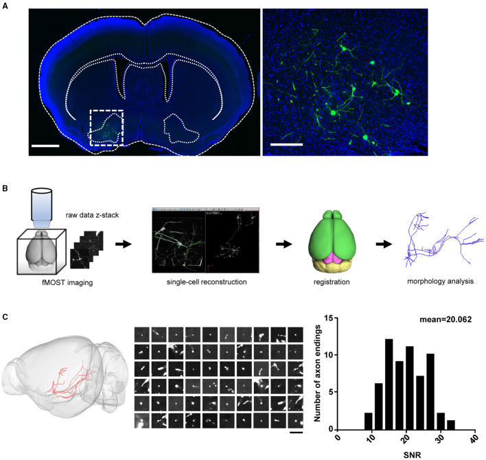
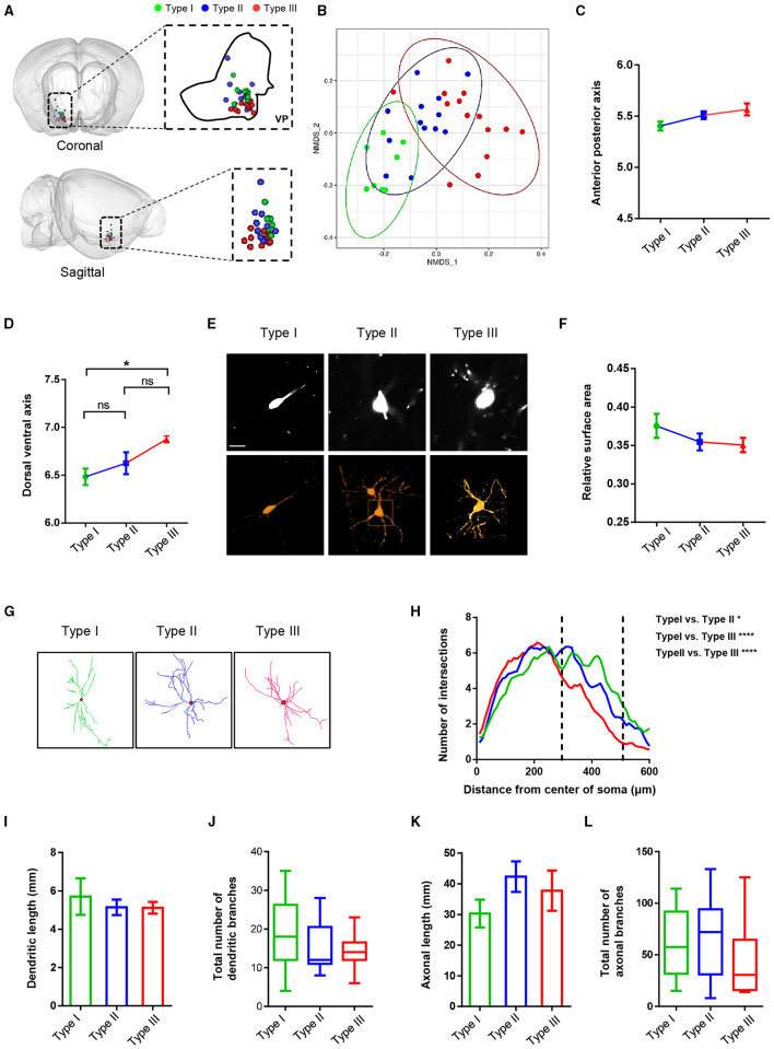
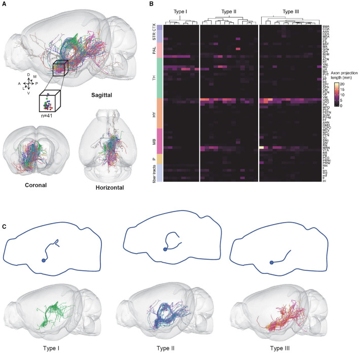
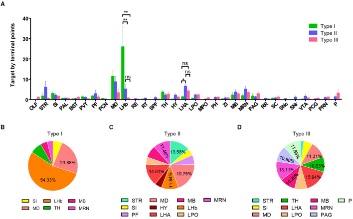
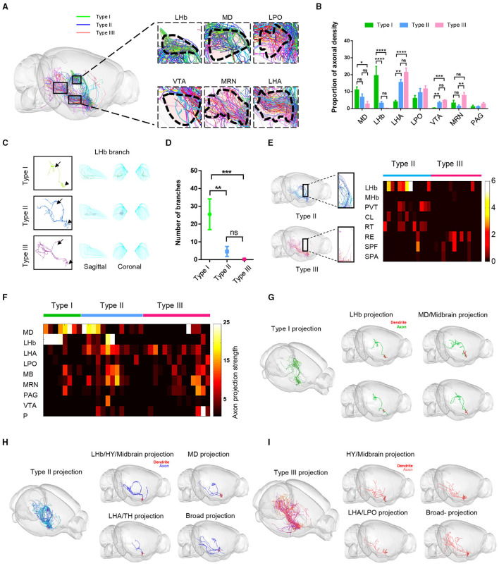
The ventral pallidum (VP) integrates reward signals to regulate cognitive, emotional, and motor processes associated with motivational salience. Previous studies have revealed that the VP projects axons to many cortical and subcortical structures. However, descriptions of the neuronal morphologies and projection patterns of the VP neurons at the single neuron level are lacking, thus hindering the understanding of the wiring diagram of the VP. In this study, we used recently developed progress in robust sparse labeling and fluorescence micro-optical sectioning tomography imaging system (fMOST) to label mediodorsal thalamus-projecting neurons in the VP and obtain high-resolution whole-brain imaging data. Based on these data, we reconstructed VP neurons and classified them into three types according to their fiber projection patterns. We systematically compared the axonal density in various downstream centers and analyzed the soma distribution and dendritic morphologies of the various subtypes at the single neuron level. Our study thus provides a detailed characterization of the morphological features of VP neurons, laying a foundation for exploring the neural circuit organization underlying the important behavioral functions of VP.
腹侧苍白球(VP)整合奖赏信号,以调节与动机显著性相关的认知、情感和运动过程。先前的研究表明,VP向许多皮质和皮质下结构投射轴突。然而,目前缺乏对VP神经元在单神经元水平上的神经元形态和投射模式的描述,这阻碍了对VP布线图的理解。在本研究中,我们利用最近在稳健稀疏标记和荧光显微光学切片断层成像系统(fMOST)方面取得的进展,标记VP中投射到丘脑背内侧核的神经元,并获得高分辨率全脑成像数据。基于这些数据,我们重建了VP神经元,并根据其纤维投射模式将它们分为三种类型。我们系统地比较了各个下游中枢的轴突密度,并在单神经元水平分析了各种亚型的胞体分布和树突形态。因此,我们的研究详细描述了VP神经元的形态特征,为探索VP重要行为功能背后的神经回路组织奠定了基础。