Kim Alex Ju Sung, Ong Sungmoon, Kim Ji Hyun, Lee Hong Sub, Yoon Jun Sik, Hur Dae Young
Department of Anatomy and Tumor Immunology, Inje University College of Medicine, Busan, Korea.
Department of Internal Medicine, Busan Paik Hospital, Inje University College of Medicine, Busan, Korea.
J Neurogastroenterol Motil. 2022 Jan 30;28(1):131-144. doi: 10.5056/jnm21020.
BACKGROUND/AIMS: The effect of peroral endoscopic myotomy (POEM) on esophageal body movement in achalasia is poorly understood. This study aims to evaluate morphological changes in esophageal body movement after POEM in type III achalasia by analyzing intraluminal ultrasound (US) images in comparison to type I and II achalasia.
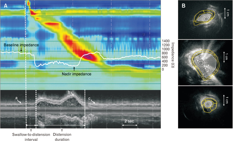
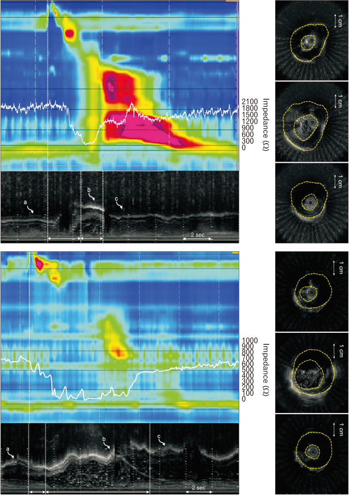
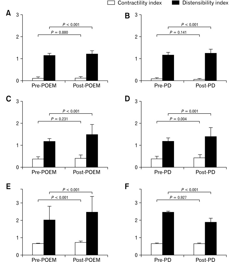
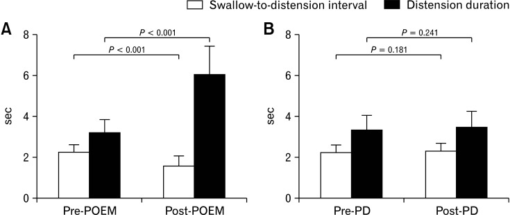
Intraluminal US images and impedance values of the distal esophagus from 47 achalasia patients who underwent POEM or pneumatic dilatation (PD) (30 patients in the POEM group and 17 patients in the PD group) with pre- and post-procedural high-resolution impedance manometry and intraluminal US examinations were analyzed. The muscle thickness (MT), muscle cross-sectional area, lumen cross-sectional area (LCSA), contractility and distensibility indices, swallow-to-distension interval, and distension duration during each bolus transport were analyzed.
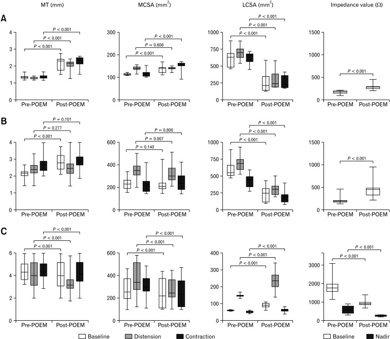
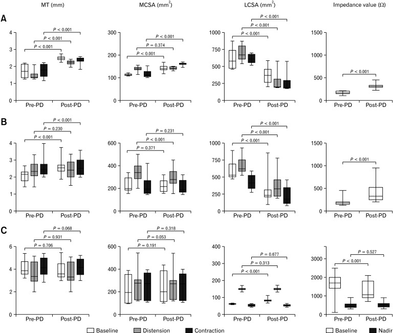
The MT increased and LCSA decreased significantly ( < 0.001), but the contractility index was not improved after POEM or PD in type I achalasia. Baseline MT increased and LCSA decreased significantly after POEM and PD in type II achalasia ( < 0.001). In contrast, MT and the swallow-to-distension interval decreased and the distension LCSA/duration and contractility index increased after POEM in type III achalasia ( < 0.001). In contrast to type I and II achalasia, in type III achalasia, these effects were unique to the POEM group.
POEM decreased the esophageal LCSA by decreasing intrabolus pressure without improving contractility in type I and II achalasia. In contrast, POEM increased esophageal body distension and contractility and improved the inhibitory process during bolus transport in type III achalasia.
背景/目的:经口内镜下肌切开术(POEM)对贲门失弛缓症食管体部运动的影响尚不清楚。本研究旨在通过分析腔内超声(US)图像,评估III型贲门失弛缓症患者在POEM术后食管体部运动的形态学变化,并与I型和II型贲门失弛缓症进行比较。
分析47例接受POEM或气囊扩张术(PD)的贲门失弛缓症患者(POEM组30例,PD组17例)术前和术后的高分辨率阻抗测压及腔内超声检查的腔内US图像和食管远端阻抗值。分析每次食团运输过程中的肌肉厚度(MT)、肌肉横截面积、管腔横截面积(LCSA)、收缩性和扩张性指数、吞咽至扩张间隔以及扩张持续时间。
I型贲门失弛缓症患者在POEM或PD术后,MT增加,LCSA显著减小(<0.001),但收缩性指数未改善。II型贲门失弛缓症患者在POEM和PD术后,基线MT增加,LCSA显著减小(<0.001)。相比之下,III型贲门失弛缓症患者在POEM术后,MT和吞咽至扩张间隔减小,扩张LCSA/持续时间和收缩性指数增加(<0.001)。与I型和II型贲门失弛缓症不同,在III型贲门失弛缓症中,这些效应仅在POEM组中出现。
在I型和II型贲门失弛缓症中,POEM通过降低食团内压力降低食管LCSA,但未改善收缩性。相比之下,在III型贲门失弛缓症中,POEM增加了食管体部的扩张和收缩性,并改善了食团运输过程中的抑制过程。