• 文献检索
  • 文档翻译
  • 深度研究
  • 学术资讯
  • Suppr Zotero 插件Zotero 插件
  • 邀请有礼
  • 套餐&价格
  • 历史记录
应用&插件
Suppr Zotero 插件Zotero 插件浏览器插件Mac 客户端Windows 客户端微信小程序
定价
高级版会员购买积分包购买API积分包
服务
文献检索文档翻译深度研究API 文档MCP 服务
关于我们
关于 Suppr公司介绍联系我们用户协议隐私条款
关注我们

Suppr 超能文献

核心技术专利:CN118964589B侵权必究
粤ICP备2023148730 号-1Suppr @ 2026

文献检索

告别复杂PubMed语法,用中文像聊天一样搜索,搜遍4000万医学文献。AI智能推荐,让科研检索更轻松。

立即免费搜索

文件翻译

保留排版,准确专业,支持PDF/Word/PPT等文件格式,支持 12+语言互译。

免费翻译文档

深度研究

AI帮你快速写综述,25分钟生成高质量综述,智能提取关键信息,辅助科研写作。

立即免费体验

贲门失弛缓症患者经 POEM 治疗的食管自动三维重建:评估治疗效果的一种创新方法。

Automatic three-dimensional reconstruction of the oesophagus in achalasia patients undergoing POEM: an innovative approach for evaluating treatment outcomes.

机构信息

Department of Gastroenterology, University Hospital Augsburg, Augsburg, Germany.

University of Augsburg, Augsburg, Germany.

出版信息

BMJ Open Gastroenterol. 2024 Jun 6;11(1):e001396. doi: 10.1136/bmjgast-2024-001396.

DOI:10.1136/bmjgast-2024-001396
PMID:38844375
原文链接:https://pmc.ncbi.nlm.nih.gov/articles/PMC11167450/
Abstract

BACKGROUND AND AIMS

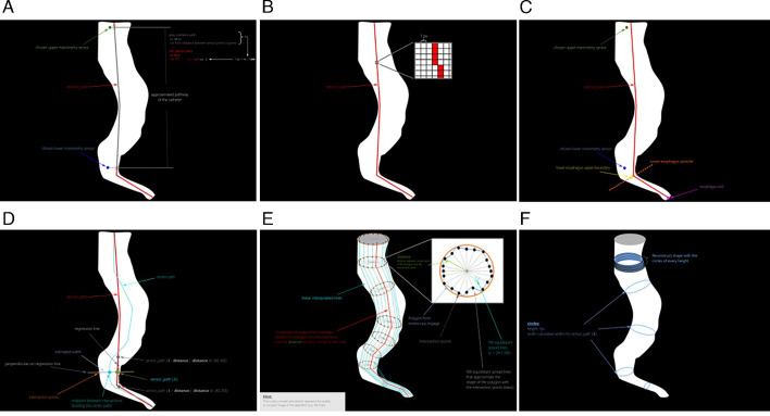
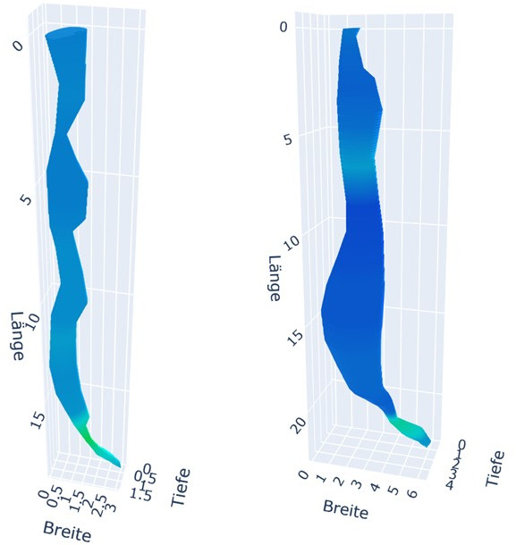
Peroral endoscopic myotomy (POEM) is a standard treatment option for achalasia patients. Treatment response varies due to factors such as achalasia type, degree of dilatation, pressure and distensibility indices. We present an innovative approach for treatment response prediction based on an automatic three-dimensional (3-D) reconstruction of the tubular oesophagus (TE) and the lower oesophageal sphincter (LES) in patients undergoing POEM for achalasia.

METHODS

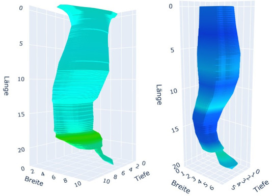
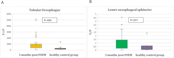
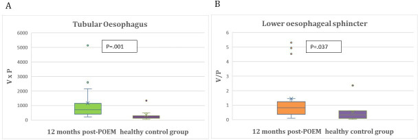
A software was developed, integrating data from high-resolution manometry, timed barium oesophagogram and endoscopic images to automatically generate 3-D reconstructions of the TE and LES. Novel normative indices for TE (volume×pressure) and LES (volume/pressure) were automatically integrated, facilitating pre-POEM and post-POEM comparisons. Treatment response was evaluated by changes in volumetric and pressure indices for the TE and the LES before as well as 3 and 12 months after POEM. In addition, these values were compared with normal value indices of non-achalasia patients.

RESULTS

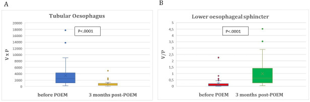
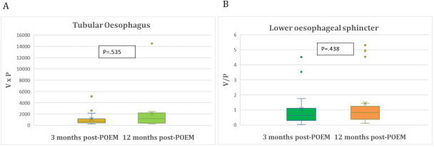
50 treatment-naive achalasia patients were enrolled prospectively. The mean TE index decreased significantly (p<0.0001) and the mean LES index increased significantly 3 months post-POEM (p<0.0001). In the 12-month follow-up, no further significant change of value indices between 3 and 12 months post-POEM was seen. 3 months post-POEM mean LES index approached the mean LES of the healthy control group (p=0.077).

CONCLUSION

3-D reconstruction provides an interactive, dynamic visualisation of the oesophagus, serving as a comprehensive tool for evaluating treatment response. It may contribute to refining our approach to achalasia treatment and optimising treatment outcomes.

TRIAL REGISTRATION NUMBER

22-0149.

摘要

背景和目的

经口内镜下肌切开术(POEM)是治疗贲门失弛缓症患者的标准治疗选择。由于贲门失弛缓症的类型、扩张程度、压力和可扩张性指数等因素,治疗反应存在差异。我们提出了一种基于贲门失弛缓症患者 POEM 治疗前后管状食管(TE)和食管下括约肌(LES)自动三维(3-D)重建的创新治疗反应预测方法。

方法

开发了一种软件,整合高分辨率测压、定时钡餐食管造影和内镜图像数据,自动生成 TE 和 LES 的 3-D 重建。自动整合 TE(容积×压力)和 LES(容积/压力)的新规范指数,便于术前和术后比较。通过 POEM 前后 TE 和 LES 的容积和压力指数变化来评估治疗反应。此外,将这些值与非贲门失弛缓症患者的正常值指数进行比较。

结果

前瞻性纳入 50 例初治贲门失弛缓症患者。TE 指数显著降低(p<0.0001),POEM 后 3 个月 LES 指数显著升高(p<0.0001)。12 个月随访时,POEM 后 3 至 12 个月之间的数值指数无进一步显著变化。POEM 后 3 个月时 LES 指数接近健康对照组的平均 LES(p=0.077)。

结论

3-D 重建提供了食管的交互式、动态可视化,是评估治疗反应的综合工具。它可能有助于完善我们的贲门失弛缓症治疗方法,优化治疗效果。

试验注册号

22-0149。

https://cdn.ncbi.nlm.nih.gov/pmc/blobs/68e1/11167450/ea2d60fd0b83/bmjgast-2024-001396f07.jpg
https://cdn.ncbi.nlm.nih.gov/pmc/blobs/68e1/11167450/dfef772bd072/bmjgast-2024-001396f01.jpg
https://cdn.ncbi.nlm.nih.gov/pmc/blobs/68e1/11167450/18584b222eb0/bmjgast-2024-001396f02.jpg
https://cdn.ncbi.nlm.nih.gov/pmc/blobs/68e1/11167450/6d58a15d399d/bmjgast-2024-001396f03.jpg
https://cdn.ncbi.nlm.nih.gov/pmc/blobs/68e1/11167450/8a22b7558884/bmjgast-2024-001396f04.jpg
https://cdn.ncbi.nlm.nih.gov/pmc/blobs/68e1/11167450/24267b3b9210/bmjgast-2024-001396f05.jpg
https://cdn.ncbi.nlm.nih.gov/pmc/blobs/68e1/11167450/5bc54cd7a5a2/bmjgast-2024-001396f06.jpg
https://cdn.ncbi.nlm.nih.gov/pmc/blobs/68e1/11167450/ea2d60fd0b83/bmjgast-2024-001396f07.jpg

相似文献

1
Automatic three-dimensional reconstruction of the oesophagus in achalasia patients undergoing POEM: an innovative approach for evaluating treatment outcomes.贲门失弛缓症患者经 POEM 治疗的食管自动三维重建:评估治疗效果的一种创新方法。
BMJ Open Gastroenterol. 2024 Jun 6;11(1):e001396. doi: 10.1136/bmjgast-2024-001396.
2
One year comprehensive prospective follow-up of achalasia patients after peroral endoscopic myotomy.经口内镜肌切开术治疗贲门失弛缓症患者一年的综合前瞻性随访。
Ann Med. 2021 Dec;53(1):2225-2233. doi: 10.1080/07853890.2021.2005253.
3
Effectiveness of peroral endoscopic myotomy in the treatment of achalasia: a pilot trial in Chinese Han population with a minimum of one-year follow-up.经口内镜下肌切开术治疗贲门失弛缓症的疗效:一项在中国汉族人群中至少随访一年的初步试验。
J Dig Dis. 2014 Jul;15(7):352-8. doi: 10.1111/1751-2980.12153.
4
Two-year follow-up for 45 patients with achalasia who underwent peroral endoscopic myotomy.45例接受经口内镜下肌切开术的贲门失弛缓症患者的两年随访。
Eur J Cardiothorac Surg. 2015 May;47(5):890-6. doi: 10.1093/ejcts/ezu320. Epub 2014 Sep 4.
5
Efficacy of peroral endoscopic myotomy vs other achalasia treatments in improving esophageal function.经口内镜下肌切开术与其他贲门失弛缓症治疗方法在改善食管功能方面的疗效比较。
World J Gastroenterol. 2016 May 28;22(20):4918-25. doi: 10.3748/wjg.v22.i20.4918.
6
A clinical study of peroral endoscopic myotomy reveals that impaired lower esophageal sphincter relaxation in achalasia is not only defined by high-resolution manometry.经口内镜下肌切开术的临床研究表明,贲门失弛缓症中食管下括约肌松弛功能障碍不仅可以通过高分辨率测压法定义。
PLoS One. 2018 Apr 2;13(4):e0195423. doi: 10.1371/journal.pone.0195423. eCollection 2018.
7
Safety and efficacy of the modified peroral endoscopic myotomy with shorter myotomy for achalasia patients: a prospective study.改良经口内镜下肌切开术缩短肌切开长度治疗贲门失弛缓症患者的安全性和有效性:一项前瞻性研究
Dis Esophagus. 2015 Nov-Dec;28(8):720-7. doi: 10.1111/dote.12280. Epub 2014 Sep 12.
8
Peroral endoscopic myotomy (POEM) for esophageal primary motility disorders: analysis of 100 consecutive patients.经口内镜下肌切开术(POEM)治疗食管原发性动力障碍:100例连续患者的分析
J Gastrointest Surg. 2015 Jan;19(1):161-70; discussion 170. doi: 10.1007/s11605-014-2610-5. Epub 2014 Sep 3.
9
Preliminary study of 1940 nm thulium laser usage in peroral endoscopic myotomy for achalasia.1940纳米铥激光在贲门失弛缓症经口内镜下肌切开术中应用的初步研究
Dis Esophagus. 2018 Feb 1;31(2). doi: 10.1093/dote/dox132.
10
Effect of peroral endoscopic myotomy on esophagogastric junction physiology in patients with achalasia.经口内镜下肌切开术对贲门失弛缓症患者食管胃结合部生理的影响。
Gastrointest Endosc. 2013 Jul;78(1):39-44. doi: 10.1016/j.gie.2013.01.006. Epub 2013 Feb 26.

本文引用的文献

1
MRI-MECH: mechanics-informed MRI to estimate esophageal health.MRI-MECH:基于力学信息的磁共振成像用于评估食管健康状况
Front Physiol. 2023 Jun 9;14:1195067. doi: 10.3389/fphys.2023.1195067. eCollection 2023.
2
Virtual disease landscape using mechanics-informed machine learning: Application to esophageal disorders.利用力学启发式机器学习进行虚拟疾病景观分析:在食管疾病中的应用。
Artif Intell Med. 2022 Dec;134:102435. doi: 10.1016/j.artmed.2022.102435. Epub 2022 Oct 31.
3
Peroral endoscopic myotomy versus pneumatic dilation in treatment-naive patients with achalasia: 5-year follow-up of a randomised controlled trial.
经口内镜下肌切开术与气囊扩张治疗原发性贲门失弛缓症的随机对照研究:5 年随访结果
Lancet Gastroenterol Hepatol. 2022 Dec;7(12):1103-1111. doi: 10.1016/S2468-1253(22)00300-4. Epub 2022 Oct 4.
4
Myotomy technique and esophageal contractility impact blown-out myotomy formation in achalasia: an in silico investigation.贲门失弛缓症中切开术技术和食管收缩力对“吹破”性切开术形成的影响:一项计算机模拟研究。
Am J Physiol Gastrointest Liver Physiol. 2022 May 1;322(5):G500-G512. doi: 10.1152/ajpgi.00281.2021. Epub 2022 Feb 16.
5
Morphologic Changes in Esophageal Body Movement During Bolus Transport After Peroral Endoscopic Myotomy in Type III Achalasia.III型贲门失弛缓症经口内镜下肌切开术后食团运输过程中食管体部运动的形态学变化
J Neurogastroenterol Motil. 2022 Jan 30;28(1):131-144. doi: 10.5056/jnm21020.
6
Comparison of functional lumen imaging probe and high-resolution manometry to assess response after peroral endoscopic myotomy.功能性管腔成像探头与高分辨率测压法评估经口内镜下肌切开术后反应的比较
Gastrointest Endosc. 2022 May;95(5):855-863. doi: 10.1016/j.gie.2021.12.029. Epub 2022 Jan 1.
7
Prediction of Esophageal Retention: A Study Comparing High-Resolution Manometry and Functional Luminal Imaging Probe Panometry.食管滞留预测:高分辨率测压与腔内功能成像探头测压的对比研究。
Am J Gastroenterol. 2021 Oct 1;116(10):2032-2041. doi: 10.14309/ajg.0000000000001402.
8
Mechanics informed fluoroscopy of esophageal transport.食管传输的力学透视研究。
Biomech Model Mechanobiol. 2021 Jun;20(3):925-940. doi: 10.1007/s10237-021-01420-0. Epub 2021 Mar 2.
9
Esophageal motility disorders on high-resolution manometry: Chicago classification version 4.0.高分辨率食管动力障碍:芝加哥分类版本 4.0。
Neurogastroenterol Motil. 2021 Jan;33(1):e14058. doi: 10.1111/nmo.14058.
10
Assessment of esophageal body peristaltic work using functional lumen imaging probe panometry.使用功能腔成像探头压力测量法评估食管体蠕动功能。
Am J Physiol Gastrointest Liver Physiol. 2021 Feb 1;320(2):G217-G226. doi: 10.1152/ajpgi.00324.2020. Epub 2020 Nov 11.