• 文献检索
  • 文档翻译
  • 深度研究
  • 学术资讯
  • Suppr Zotero 插件Zotero 插件
  • 邀请有礼
  • 套餐&价格
  • 历史记录
应用&插件
Suppr Zotero 插件Zotero 插件浏览器插件Mac 客户端Windows 客户端微信小程序
定价
高级版会员购买积分包购买API积分包
服务
文献检索文档翻译深度研究API 文档MCP 服务
关于我们
关于 Suppr公司介绍联系我们用户协议隐私条款
关注我们

Suppr 超能文献

核心技术专利:CN118964589B侵权必究
粤ICP备2023148730 号-1Suppr @ 2026

文献检索

告别复杂PubMed语法,用中文像聊天一样搜索,搜遍4000万医学文献。AI智能推荐,让科研检索更轻松。

立即免费搜索

文件翻译

保留排版,准确专业,支持PDF/Word/PPT等文件格式,支持 12+语言互译。

免费翻译文档

深度研究

AI帮你快速写综述,25分钟生成高质量综述,智能提取关键信息,辅助科研写作。

立即免费体验

扩散加权磁共振成像鉴别良性与恶性非脂肪性肌肉骨骼软组织肿瘤的诊断性能:一项系统评价和荟萃分析

Diagnostic Performance of Diffusion MRI for differentiating Benign and Malignant Nonfatty Musculoskeletal Soft Tissue Tumors: A Systematic Review and Meta-analysis.

作者信息

Wang Qian, Xiao Xinguang, Liang Yanchang, Wen Hao, Wen Xiaopeng, Gu Meilan, Ren Cuiping, Li Kunbin, Yu Liangwen, Lu Liming

机构信息

Department of Medical Imaging, Zhengzhou Central Hospital Affiliated to Zhengzhou University, 195 Tongbai Road, 455007, Zhengzhou, China.

Guangzhou University of Chinese Medicine, 510006, Guangzhou, China.

出版信息

J Cancer. 2021 Oct 28;12(24):7399-7412. doi: 10.7150/jca.62131. eCollection 2021.

DOI:10.7150/jca.62131
PMID:35003360
原文链接:https://pmc.ncbi.nlm.nih.gov/articles/PMC8734420/
Abstract

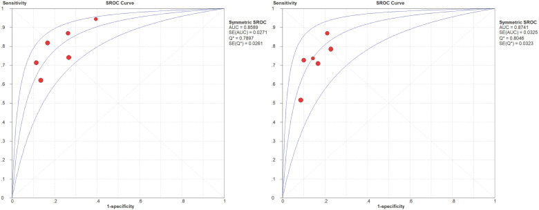
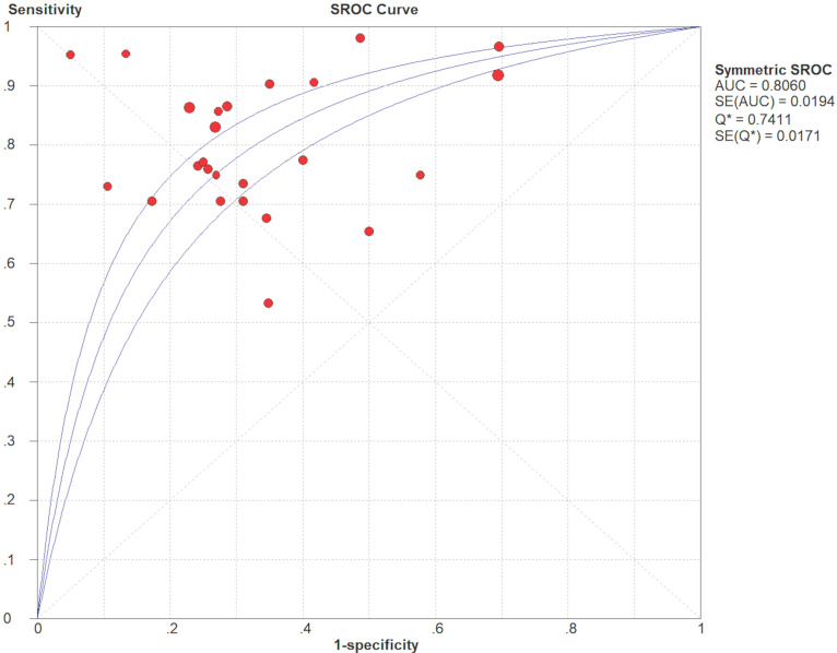
To evaluate the diagnostic performance of standard diffusion-weighted imaging (DWI), intravoxel incoherent motion (IVIM), and diffusion kurtosis imaging (DKI), for differentiating benign and malignant soft tissue tumors (STTs). A thorough search was carried out to identify suitable studies published up to September 2020. The quality of the studies involved was evaluated using Quality Assessment of Diagnostic Accuracy Studies-2 (QUADAS-2). The pooled sensitivity (SEN), specificity (SPE), and summary receiver operating characteristic (SROC) curve were calculated using bivariate mixed effects models. A subgroup analysis was also performed to explore the heterogeneity. Eighteen studies investigating 1319 patients with musculoskeletal STTs (malignant, =623; benign, =696) were enrolled. Thirteen standard DWI studies using the apparent diffusion coefficient (ADC) showed that the pooled SEN and SPE of ADC were 0.80 (95% CI: 0.77-0.82) and 0.63 (95% CI: 0.60-0.67), respectively. The area under the curve (AUC) calculated from the SROC curve was 0.806. The subgroup analysis indicated that the percentage of myxoid malignant tumors, magnet strength, study design, and ROI placement were significant factors affecting heterogeneity. Four IVIM studies showed that the AUCs calculated from the SROC curves of the parameters ADC and D were 0.859 and 0.874, respectively. The AUCs for the IVIM parameters pseudo diffusion coefficient (D*) and perfusion fraction (f) calculated from the SROC curve were 0.736 and 0.573, respectively. Two DKI studies showed that the AUCs of the DKI parameter mean kurtosis (MK) were 0.97 and 0.89, respectively. The DWI-derived ADC value and the IVIM DWI-derived D value might be accurate tools for discriminating musculoskeletal STTs, especially for non-myxoid SSTs, using more than two b values, with maximal b value ranging from 600 to 800 s/mm, additionally, a high-field strength (3.0 T) optimizes the diagnostic performance.

摘要

为评估标准扩散加权成像(DWI)、体素内不相干运动(IVIM)和扩散峰度成像(DKI)在鉴别良性和恶性软组织肿瘤(STT)方面的诊断性能。进行了全面检索以识别截至2020年9月发表的合适研究。使用诊断准确性研究质量评估-2(QUADAS-2)对所涉研究的质量进行评估。使用双变量混合效应模型计算合并敏感度(SEN)、特异度(SPE)和汇总接收器操作特征(SROC)曲线。还进行了亚组分析以探讨异质性。纳入了18项研究,共1319例肌肉骨骼STT患者(恶性,=623例;良性,=696例)。13项使用表观扩散系数(ADC)的标准DWI研究表明,ADC的合并SEN和SPE分别为0.80(95%CI:0.77-0.82)和0.63(95%CI:0.60-0.67)。从SROC曲线计算的曲线下面积(AUC)为0.806。亚组分析表明,黏液样恶性肿瘤的百分比、磁体强度、研究设计和感兴趣区(ROI)放置是影响异质性的显著因素。4项IVIM研究表明,参数ADC和D的SROC曲线计算的AUC分别为0.859和0.874。从SROC曲线计算的IVIM参数伪扩散系数(D*)和灌注分数(f)的AUC分别为0.736和0.573。2项DKI研究表明,DKI参数平均峰度(MK)的AUC分别为0.97和0.89。DWI衍生的ADC值和IVIM DWI衍生的D值可能是鉴别肌肉骨骼STT的准确工具,尤其是对于非黏液样SST,使用两个以上的b值,最大b值范围为600至800 s/mm,此外,高场强(3.0 T)可优化诊断性能。

https://cdn.ncbi.nlm.nih.gov/pmc/blobs/5013/8734420/6fb1dd891063/jcav12p7399g005.jpg
https://cdn.ncbi.nlm.nih.gov/pmc/blobs/5013/8734420/00c238badbaf/jcav12p7399g001.jpg
https://cdn.ncbi.nlm.nih.gov/pmc/blobs/5013/8734420/f2c30d3bdb95/jcav12p7399g002.jpg
https://cdn.ncbi.nlm.nih.gov/pmc/blobs/5013/8734420/804e4b7de484/jcav12p7399g003.jpg
https://cdn.ncbi.nlm.nih.gov/pmc/blobs/5013/8734420/da222d853827/jcav12p7399g004.jpg
https://cdn.ncbi.nlm.nih.gov/pmc/blobs/5013/8734420/6fb1dd891063/jcav12p7399g005.jpg

相似文献

1
Diagnostic Performance of Diffusion MRI for differentiating Benign and Malignant Nonfatty Musculoskeletal Soft Tissue Tumors: A Systematic Review and Meta-analysis.扩散加权磁共振成像鉴别良性与恶性非脂肪性肌肉骨骼软组织肿瘤的诊断性能:一项系统评价和荟萃分析
J Cancer. 2021 Oct 28;12(24):7399-7412. doi: 10.7150/jca.62131. eCollection 2021.
2
Differentiating between malignant and benign renal tumors: do IVIM and diffusion kurtosis imaging perform better than DWI?鉴别良恶性肾肿瘤:IVIM 和扩散峰度成像是否优于 DWI?
Eur Radiol. 2019 Dec;29(12):6930-6939. doi: 10.1007/s00330-019-06240-6. Epub 2019 Jun 3.
3
Differentiating between malignant and benign solid solitary pulmonary lesions: are intravoxel incoherent motion and diffusion kurtosis imaging superior to conventional diffusion-weighted imaging?鉴别良恶性肺部实性孤立性病变:体素内不相干运动和扩散峰度成像优于常规扩散加权成像吗?
Eur Radiol. 2019 Mar;29(3):1607-1615. doi: 10.1007/s00330-018-5714-6. Epub 2018 Sep 25.
4
Different Attenuation Models of Diffusion-Weighted MR Imaging for the Differentiation of Benign and Malignant Musculoskeletal Tumors.用于鉴别骨骼肌肉系统良恶性肿瘤的扩散加权磁共振成像的不同衰减模型
J Magn Reson Imaging. 2022 Feb;55(2):594-607. doi: 10.1002/jmri.27887. Epub 2021 Aug 16.
5
Intravoxel incoherent motion (IVIM) in evaluation of breast lesions: comparison with conventional DWI.体素内不相干运动(IVIM)在评估乳腺病变中的应用:与常规 DWI 的比较。
Eur J Radiol. 2013 Dec;82(12):e782-9. doi: 10.1016/j.ejrad.2013.08.006. Epub 2013 Aug 13.
6
Accuracy of quantitative diffusion-weighted imaging for differentiating benign and malignant pancreatic lesions: a systematic review and meta-analysis.定量扩散加权成像鉴别良恶性胰腺病变的准确性:系统评价和荟萃分析。
Eur Radiol. 2021 Oct;31(10):7746-7759. doi: 10.1007/s00330-021-07880-3. Epub 2021 Apr 13.
7
Comparison of Conventional DWI, Intravoxel Incoherent Motion Imaging, and Diffusion Kurtosis Imaging in Differentiating Lung Lesions.传统扩散加权成像、体素内不相干运动成像及扩散峰度成像在鉴别肺部病变中的比较
Front Oncol. 2022 Jan 20;11:815967. doi: 10.3389/fonc.2021.815967. eCollection 2021.
8
Quantitative analysis for detection and grading of hepatocellular carcinoma: Comparison of diffusion kurtosis imaging, intravoxel incoherent motion and conventional diffusion-weighted imaging.肝细胞癌检测与分级的定量分析:扩散峰度成像、体素内不相干运动与传统扩散加权成像的比较
Oncol Lett. 2022 Sep 22;24(5):403. doi: 10.3892/ol.2022.13523. eCollection 2022 Nov.
9
Diagnostic performance of diffusion-weighted imaging and intravoxel incoherent motion for renal lesions: a meta-analysis.扩散加权成像和体素内不相干运动对肾脏病变的诊断性能:荟萃分析。
Clin Radiol. 2023 Dec;78(12):935-946. doi: 10.1016/j.crad.2023.07.024. Epub 2023 Aug 19.
10
Intravoxel incoherent motion diffusion-weighted MR imaging for differentiation of benign and malignant musculoskeletal tumours at 3 T.3T磁共振体素内不相干运动扩散加权成像用于鉴别骨骼肌肉系统良恶性肿瘤
Br J Radiol. 2018 Feb;91(1082):20170636. doi: 10.1259/bjr.20170636. Epub 2017 Dec 15.

引用本文的文献

1
Recent Advances in Magnetic Resonance Imaging for the Diagnosis of Liver Cancer: A Comprehensive Review.磁共振成像在肝癌诊断中的最新进展:综述
Diagnostics (Basel). 2025 Aug 12;15(16):2016. doi: 10.3390/diagnostics15162016.
2
Whole-tumor histogram analysis of synthetic MRI for the differentiation of benign and malignant soft-tissue tumors: a preliminary study.基于合成磁共振成像的全肿瘤直方图分析在鉴别良恶性软组织肿瘤中的应用:一项初步研究
Eur Radiol. 2025 Jun 26. doi: 10.1007/s00330-025-11770-3.
3
Comparison of diagnostic performance between diffusion models parameters and mono-exponential apparent diffusion coefficient in patients with prostate cancer: A systematic review and meta-analysis.

本文引用的文献

1
Revisiting the WHO classification system of soft tissue tumours: emphasis on advanced magnetic resonance imaging sequences. Part 1.重新审视世界卫生组织软组织肿瘤分类系统:重点关注先进的磁共振成像序列。第1部分。
Pol J Radiol. 2020 Aug 3;85:e396-e408. doi: 10.5114/pjr.2020.98685. eCollection 2020.
2
Added-value of advanced magnetic resonance imaging to conventional morphologic analysis for the differentiation between benign and malignant non-fatty soft-tissue tumors.高级磁共振成像对常规形态分析在鉴别良恶性非脂肪性软组织肿瘤中的附加价值。
Eur Radiol. 2021 Mar;31(3):1536-1547. doi: 10.1007/s00330-020-07190-0. Epub 2020 Sep 3.
3
前列腺癌患者中扩散模型参数与单指数表观扩散系数诊断性能的比较:一项系统评价和荟萃分析
J Res Med Sci. 2024 Jul 30;29:43. doi: 10.4103/jrms.jrms_359_23. eCollection 2024.
4
Soft tissue vascular tumor-like lesions in adults: imaging and pathological analysis pitfalls per ISSVA classification.成人软组织血管肿瘤样病变:基于国际血管性肿瘤学会(ISSVA)分类的影像学及病理分析陷阱
Insights Imaging. 2024 Jun 9;15(1):135. doi: 10.1186/s13244-024-01712-w.
5
Differentiating nasopharyngeal carcinoma from lymphoma in the head and neck region using the apparent diffusion coefficient (ADC) value: a systematic review and meta-analysis.利用表观扩散系数(ADC)值鉴别头颈部区域的鼻咽癌与淋巴瘤:一项系统评价和荟萃分析。
Pol J Radiol. 2023 Oct 17;88:e472-e482. doi: 10.5114/pjr.2023.132172. eCollection 2023.
6
Pitfalls of Diffusion-Weighted Imaging: Clinical Utility of T2 Shine-through and T2 Black-out for Musculoskeletal Diseases.扩散加权成像的陷阱:T2 透过效应和 T2 信号缺失在肌肉骨骼疾病中的临床应用
Diagnostics (Basel). 2023 May 7;13(9):1647. doi: 10.3390/diagnostics13091647.
7
Diffusion-Weighted Magnetic Resonance Imaging: Clinical Potential and Applications.扩散加权磁共振成像:临床潜力与应用
J Clin Med. 2022 Jun 10;11(12):3339. doi: 10.3390/jcm11123339.
8
Features of ophthalmic, magnetic resonance imaging, and histopathology of a feline case of idiopathic sclerosing orbital pseudotumor.一例特发性硬化性眼眶假瘤猫的眼科、磁共振成像和组织病理学特征。
Vet Med Sci. 2022 Jul;8(4):1352-1360. doi: 10.1002/vms3.822. Epub 2022 May 3.
Multiparametric quantitative analysis of tumor perfusion and diffusion with 3T MRI: differentiation between benign and malignant soft tissue tumors.
3T MRI 多参数定量分析肿瘤灌注和弥散:鉴别良恶性软组织肿瘤。
Br J Radiol. 2020 Nov 1;93(1115):20191035. doi: 10.1259/bjr.20191035. Epub 2020 Oct 1.
4
Characterization of small, deeply located soft-tissue tumors: Conventional magnetic resonance imaging features and apparent diffusion coefficient for differentiation between non-malignancy and malignancy.小而深在的软组织肿瘤的特征:常规磁共振成像特征与表观扩散系数在良恶性鉴别诊断中的应用。
PLoS One. 2020 May 7;15(5):e0232622. doi: 10.1371/journal.pone.0232622. eCollection 2020.
5
Intravoxel incoherent motion MRI for the initial characterization of non-fatty non-vascular soft tissue tumors.体素内不相干运动 MRI 用于非脂肪性非血管性软组织肿瘤的初步特征描述。
Diagn Interv Imaging. 2020 Apr;101(4):245-255. doi: 10.1016/j.diii.2019.11.003. Epub 2019 Dec 11.
6
Advanced MR Imaging of Musculoskeletal Tumors: An Overview.肌肉骨骼肿瘤的高级磁共振成像:概述
Semin Roentgenol. 2019 Apr;54(2):149-161. doi: 10.1053/j.ro.2018.10.001. Epub 2018 Oct 10.
7
A Meta-analysis of the Diagnostic Performance of Diffusion MRI for Breast Lesion Characterization.磁共振弥散成像对乳腺病变特征诊断性能的 Meta 分析。
Radiology. 2019 Jun;291(3):632-641. doi: 10.1148/radiol.2019182510. Epub 2019 Apr 23.
8
Advanced magnetic resonance imaging (MRI) of soft tissue tumors: techniques and applications.软组织肿瘤的高级磁共振成像(MRI):技术与应用。
Radiol Med. 2019 Apr;124(4):243-252. doi: 10.1007/s11547-019-01035-7. Epub 2019 Apr 4.
9
Diagnostic performance of DWI for differentiating primary central nervous system lymphoma from glioblastoma: a systematic review and meta-analysis.DWI 对原发性中枢神经系统淋巴瘤与胶质母细胞瘤鉴别诊断的性能:系统评价和荟萃分析。
Neurol Sci. 2019 May;40(5):947-956. doi: 10.1007/s10072-019-03732-7. Epub 2019 Jan 31.
10
Diagnostic performance of diffusion-weighted (DWI) and dynamic contrast-enhanced (DCE) MRI for the differentiation of benign from malignant soft-tissue tumors.弥散加权(DWI)和动态对比增强(DCE)MRI 对良恶性软组织肿瘤的鉴别诊断性能。
J Magn Reson Imaging. 2019 Sep;50(3):798-809. doi: 10.1002/jmri.26607. Epub 2019 Jan 20.