• 文献检索
  • 文档翻译
  • 深度研究
  • 学术资讯
  • Suppr Zotero 插件Zotero 插件
  • 邀请有礼
  • 套餐&价格
  • 历史记录
应用&插件
Suppr Zotero 插件Zotero 插件浏览器插件Mac 客户端Windows 客户端微信小程序
定价
高级版会员购买积分包购买API积分包
服务
文献检索文档翻译深度研究API 文档MCP 服务
关于我们
关于 Suppr公司介绍联系我们用户协议隐私条款
关注我们

Suppr 超能文献

核心技术专利:CN118964589B侵权必究
粤ICP备2023148730 号-1Suppr @ 2026

文献检索

告别复杂PubMed语法,用中文像聊天一样搜索,搜遍4000万医学文献。AI智能推荐,让科研检索更轻松。

立即免费搜索

文件翻译

保留排版,准确专业,支持PDF/Word/PPT等文件格式,支持 12+语言互译。

免费翻译文档

深度研究

AI帮你快速写综述,25分钟生成高质量综述,智能提取关键信息,辅助科研写作。

立即免费体验

将心肌细胞增殖作为促进心脏损伤后修复的关键方法。

Targeting cardiomyocyte proliferation as a key approach of promoting heart repair after injury.

作者信息

Li Shuainan, Ma Wenya, Cai Benzhi

机构信息

Department of Pharmacy at The Second Affiliated Hospital, and Department of Pharmacology at College of Pharmacy (The Key Laboratory of Cardiovascular Medicine Research, Ministry of Education), Harbin Medical University, Harbin, 150086, China.

Institute of Clinical Pharmacy, the Heilongjiang Key Laboratory of Drug Research, Harbin Medical University, Harbin, 150086, China.

出版信息

Mol Biomed. 2021 Nov 5;2(1):34. doi: 10.1186/s43556-021-00047-y.

DOI:10.1186/s43556-021-00047-y
PMID:35006441
原文链接:https://pmc.ncbi.nlm.nih.gov/articles/PMC8607366/
Abstract

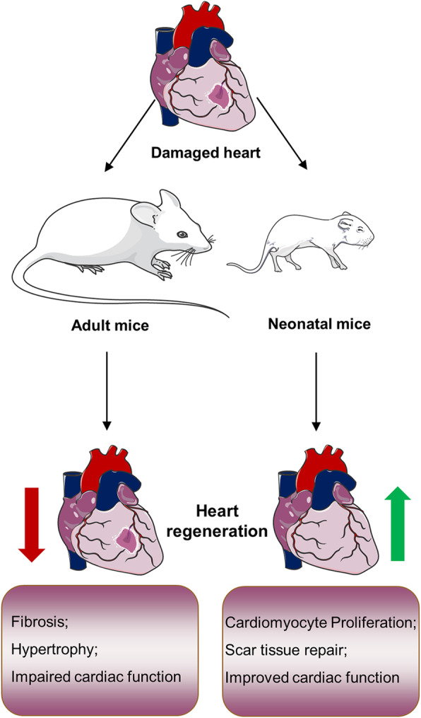
Cardiovascular diseases such as myocardial infarction (MI) is a major contributor to human mortality and morbidity. The mammalian adult heart almost loses its plasticity to appreciably regenerate new cardiomyocytes after injuries, such as MI and heart failure. The neonatal heart exhibits robust proliferative capacity when exposed to varying forms of myocardial damage. The ability of the neonatal heart to repair the injury and prevent pathological left ventricular remodeling leads to preserved or improved cardiac function. Therefore, promoting cardiomyocyte proliferation after injuries to reinitiate the process of cardiomyocyte regeneration, and suppress heart failure and other serious cardiovascular problems have become the primary goal of many researchers. Here, we review recent studies in this field and summarize the factors that act upon the proliferation of cardiomyocytes and cardiac repair after injury and discuss the new possibilities for potential clinical treatment strategies for cardiovascular diseases.

摘要

心血管疾病,如心肌梗死(MI),是导致人类死亡和发病的主要因素。成年哺乳动物的心脏在遭受诸如心肌梗死和心力衰竭等损伤后,几乎失去了显著再生新心肌细胞的可塑性。新生儿心脏在受到不同形式的心肌损伤时,表现出强大的增殖能力。新生儿心脏修复损伤和预防病理性左心室重塑的能力可维持或改善心脏功能。因此,促进损伤后心肌细胞增殖以重新启动心肌细胞再生过程,抑制心力衰竭和其他严重心血管问题,已成为许多研究人员的首要目标。在此,我们综述了该领域的最新研究,总结了影响损伤后心肌细胞增殖和心脏修复的因素,并探讨了心血管疾病潜在临床治疗策略的新可能性。

https://cdn.ncbi.nlm.nih.gov/pmc/blobs/f3d8/8607366/63483be60f14/43556_2021_47_Fig4_HTML.jpg
https://cdn.ncbi.nlm.nih.gov/pmc/blobs/f3d8/8607366/97302dbdafb2/43556_2021_47_Fig1_HTML.jpg
https://cdn.ncbi.nlm.nih.gov/pmc/blobs/f3d8/8607366/e4e70c9d8c6e/43556_2021_47_Fig2_HTML.jpg
https://cdn.ncbi.nlm.nih.gov/pmc/blobs/f3d8/8607366/33223bb6ab7c/43556_2021_47_Fig3_HTML.jpg
https://cdn.ncbi.nlm.nih.gov/pmc/blobs/f3d8/8607366/63483be60f14/43556_2021_47_Fig4_HTML.jpg

相似文献

1
Targeting cardiomyocyte proliferation as a key approach of promoting heart repair after injury.将心肌细胞增殖作为促进心脏损伤后修复的关键方法。
Mol Biomed. 2021 Nov 5;2(1):34. doi: 10.1186/s43556-021-00047-y.
2
Regulation of cardiomyocyte fate plasticity: a key strategy for cardiac regeneration.调控心肌细胞命运可塑性:心脏再生的关键策略。
Signal Transduct Target Ther. 2021 Jan 27;6(1):31. doi: 10.1038/s41392-020-00413-2.
3
Mydgf promotes Cardiomyocyte proliferation and Neonatal Heart regeneration.Mydgf 促进心肌细胞增殖和新生儿心脏再生。
Theranostics. 2020 Jul 11;10(20):9100-9112. doi: 10.7150/thno.44281. eCollection 2020.
4
Long Noncoding RNA CPR (Cardiomyocyte Proliferation Regulator) Regulates Cardiomyocyte Proliferation and Cardiac Repair.长链非编码 RNA CPR(心肌细胞增殖调节剂)调节心肌细胞增殖和心脏修复。
Circulation. 2019 Jun 4;139(23):2668-2684. doi: 10.1161/CIRCULATIONAHA.118.035832. Epub 2019 Mar 5.
5
MicroRNA-34a Plays a Key Role in Cardiac Repair and Regeneration Following Myocardial Infarction.微小RNA-34a在心肌梗死后的心脏修复和再生中起关键作用。
Circ Res. 2015 Aug 14;117(5):450-9. doi: 10.1161/CIRCRESAHA.117.305962. Epub 2015 Jun 16.
6
Manipulating Cardiomyocyte Plasticity for Heart Regeneration.通过调控心肌细胞可塑性实现心脏再生
Front Cell Dev Biol. 2022 Jul 11;10:929256. doi: 10.3389/fcell.2022.929256. eCollection 2022.
7
Decellularized neonatal cardiac extracellular matrix prevents widespread ventricular remodeling in adult mammals after myocardial infarction.去细胞化的新生心脏细胞外基质可预防成年哺乳动物心肌梗死后的广泛心室重构。
Acta Biomater. 2019 Mar 15;87:140-151. doi: 10.1016/j.actbio.2019.01.062. Epub 2019 Jan 30.
8
gp130 Controls Cardiomyocyte Proliferation and Heart Regeneration.gp130调控心肌细胞增殖与心脏再生。
Circulation. 2020 Sep 8;142(10):967-982. doi: 10.1161/CIRCULATIONAHA.119.044484. Epub 2020 Jun 30.
9
LRP6 downregulation promotes cardiomyocyte proliferation and heart regeneration.LRP6 下调促进心肌细胞增殖和心脏再生。
Cell Res. 2021 Apr;31(4):450-462. doi: 10.1038/s41422-020-00411-7. Epub 2020 Sep 24.
10
Cardiomyocyte death and renewal in the normal and diseased heart.正常及患病心脏中的心肌细胞死亡与更新
Cardiovasc Pathol. 2008 Nov-Dec;17(6):349-74. doi: 10.1016/j.carpath.2008.02.004. Epub 2008 Apr 1.

引用本文的文献

1
Crosstalk between MST1-Hippo and Wnt/β-Catenin, Notch, and PI3K/Akt pathways in cardiac physiology and pathology.MST1-河马通路与Wnt/β-连环蛋白、Notch和PI3K/Akt通路在心脏生理和病理过程中的相互作用。
J Physiol Biochem. 2025 Jun 9. doi: 10.1007/s13105-025-01099-2.
2
RNA-Binding Proteins as Critical Post-Transcriptional Regulators of Cardiac Regeneration.RNA结合蛋白作为心脏再生的关键转录后调节因子
Int J Mol Sci. 2023 Jul 26;24(15):12004. doi: 10.3390/ijms241512004.
3
Signs and signals limiting myocardial damage using PICSO: a scoping review decoding paradigm shifts toward a new encounter.

本文引用的文献

1
Krüppel-like factor 1 is a core cardiomyogenic trigger in zebrafish.Krüppel-like factor 1 是斑马鱼心脏发生的核心触发因子。
Science. 2021 Apr 9;372(6538):201-205. doi: 10.1126/science.abe2762.
2
Engineering Extracellular Matrix Proteins to Enhance Cardiac Regeneration After Myocardial Infarction.工程化细胞外基质蛋白以增强心肌梗死后的心脏再生
Front Bioeng Biotechnol. 2021 Jan 20;8:611936. doi: 10.3389/fbioe.2020.611936. eCollection 2020.
3
The FGF-AKT pathway is necessary for cardiomyocyte survival for heart regeneration in zebrafish.
使用压力增强型心冲击振荡法限制心肌损伤的体征和信号:一项范围综述解码向新情况的范式转变。
Front Cardiovasc Med. 2023 May 9;10:1030842. doi: 10.3389/fcvm.2023.1030842. eCollection 2023.
4
IGF2BP3 promotes adult myocardial regeneration by stabilizing MMP3 mRNA through interaction with m6A modification.IGF2BP3通过与m6A修饰相互作用稳定MMP3 mRNA,从而促进成年心肌再生。
Cell Death Discov. 2023 May 15;9(1):164. doi: 10.1038/s41420-023-01457-3.
FGF-AKT 通路对于斑马鱼心脏再生中的心肌细胞存活是必需的。
Dev Biol. 2021 Apr;472:30-37. doi: 10.1016/j.ydbio.2020.12.019. Epub 2021 Jan 11.
4
Hypoxia-induced amniotic fluid stem cell secretome augments cardiomyocyte proliferation and enhances cardioprotective effects under hypoxic-ischemic conditions.缺氧诱导的羊水干细胞分泌组增强心肌细胞增殖,并在缺氧缺血条件下增强心脏保护作用。
Sci Rep. 2021 Jan 8;11(1):163. doi: 10.1038/s41598-020-80326-w.
5
Effect of cellular and ECM aging on human iPSC-derived cardiomyocyte performance, maturity and senescence.细胞和细胞外基质衰老对人诱导多能干细胞源性心肌细胞功能、成熟和衰老的影响。
Biomaterials. 2021 Jan;268:120554. doi: 10.1016/j.biomaterials.2020.120554. Epub 2020 Nov 22.
6
β-Catenin drives distinct transcriptional networks in proliferative and nonproliferative cardiomyocytes.β-连环蛋白在增殖型和非增殖型心肌细胞中驱动不同的转录网络。
Development. 2020 Nov 30;147(22):dev193417. doi: 10.1242/dev.193417.
7
ERBB2 drives YAP activation and EMT-like processes during cardiac regeneration.ERBB2 驱动心脏再生过程中的 YAP 激活和 EMT 样过程。
Nat Cell Biol. 2020 Nov;22(11):1346-1356. doi: 10.1038/s41556-020-00588-4. Epub 2020 Oct 12.
8
Tp53 Suppression Promotes Cardiomyocyte Proliferation during Zebrafish Heart Regeneration.抑癌基因 TP53 促进斑马鱼心脏再生过程中心肌细胞的增殖。
Cell Rep. 2020 Sep 1;32(9):108089. doi: 10.1016/j.celrep.2020.108089.
9
Tbx20 Induction Promotes Zebrafish Heart Regeneration by Inducing Cardiomyocyte Dedifferentiation and Endocardial Expansion.Tbx20诱导通过诱导心肌细胞去分化和心内膜扩张促进斑马鱼心脏再生。
Front Cell Dev Biol. 2020 Aug 4;8:738. doi: 10.3389/fcell.2020.00738. eCollection 2020.
10
Mydgf promotes Cardiomyocyte proliferation and Neonatal Heart regeneration.Mydgf 促进心肌细胞增殖和新生儿心脏再生。
Theranostics. 2020 Jul 11;10(20):9100-9112. doi: 10.7150/thno.44281. eCollection 2020.