• 文献检索
  • 文档翻译
  • 深度研究
  • 学术资讯
  • Suppr Zotero 插件Zotero 插件
  • 邀请有礼
  • 套餐&价格
  • 历史记录
应用&插件
Suppr Zotero 插件Zotero 插件浏览器插件Mac 客户端Windows 客户端微信小程序
定价
高级版会员购买积分包购买API积分包
服务
文献检索文档翻译深度研究API 文档MCP 服务
关于我们
关于 Suppr公司介绍联系我们用户协议隐私条款
关注我们

Suppr 超能文献

核心技术专利:CN118964589B侵权必究
粤ICP备2023148730 号-1Suppr @ 2026

文献检索

告别复杂PubMed语法,用中文像聊天一样搜索,搜遍4000万医学文献。AI智能推荐,让科研检索更轻松。

立即免费搜索

文件翻译

保留排版,准确专业,支持PDF/Word/PPT等文件格式,支持 12+语言互译。

免费翻译文档

深度研究

AI帮你快速写综述,25分钟生成高质量综述,智能提取关键信息,辅助科研写作。

立即免费体验

抑癌基因 TP53 促进斑马鱼心脏再生过程中心肌细胞的增殖。

Tp53 Suppression Promotes Cardiomyocyte Proliferation during Zebrafish Heart Regeneration.

机构信息

Regeneration Next, Duke University, Durham, NC 27710, USA; Department of Surgery, Duke University Medical Center, Durham, NC 27710, USA; Department of Cell Biology, Duke University Medical Center, Durham, NC 27710, USA.

Regeneration Next, Duke University, Durham, NC 27710, USA; Department of Cell Biology, Duke University Medical Center, Durham, NC 27710, USA.

出版信息

Cell Rep. 2020 Sep 1;32(9):108089. doi: 10.1016/j.celrep.2020.108089.

DOI:10.1016/j.celrep.2020.108089
PMID:32877671
原文链接:https://pmc.ncbi.nlm.nih.gov/articles/PMC7494019/
Abstract

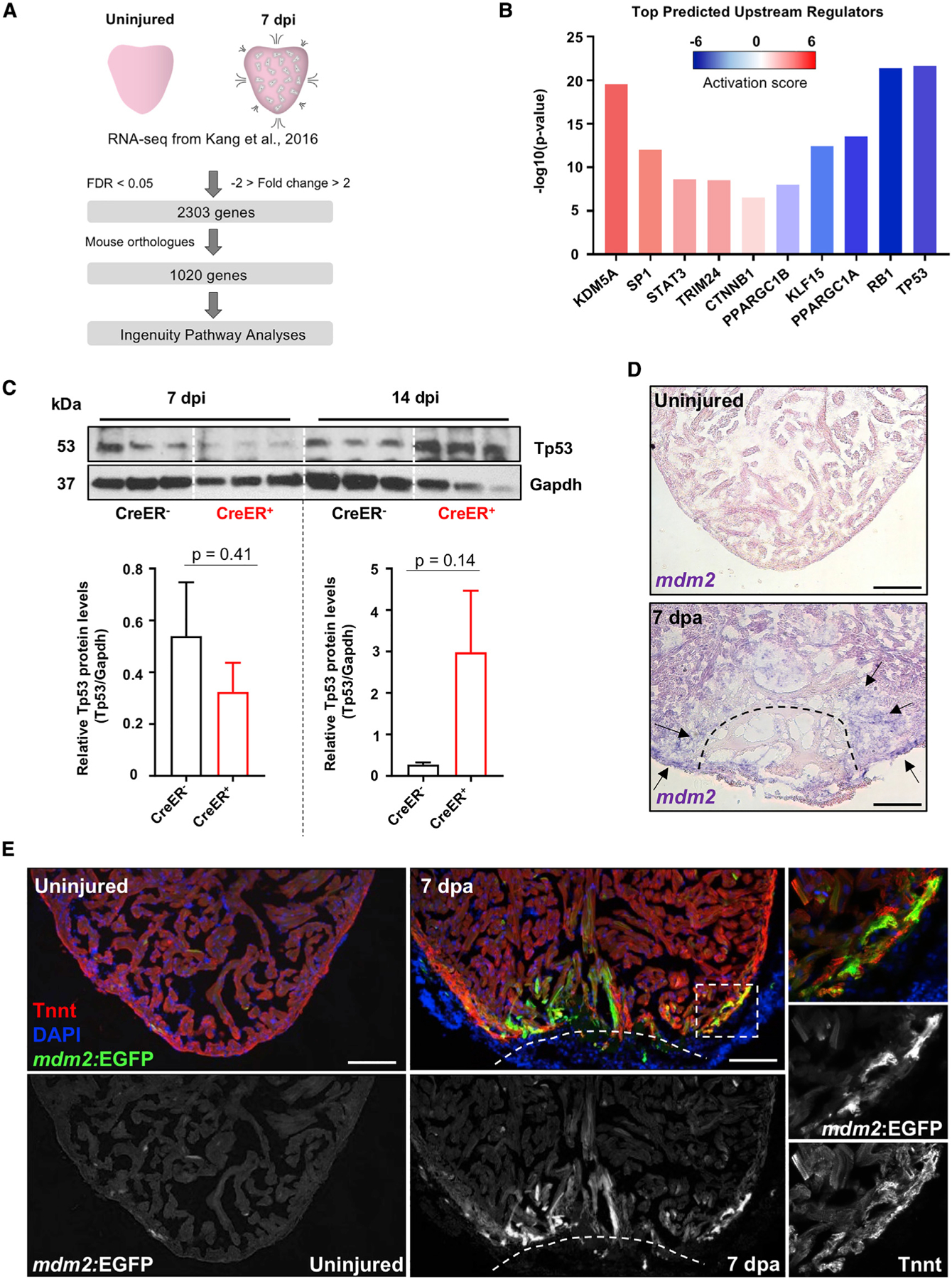
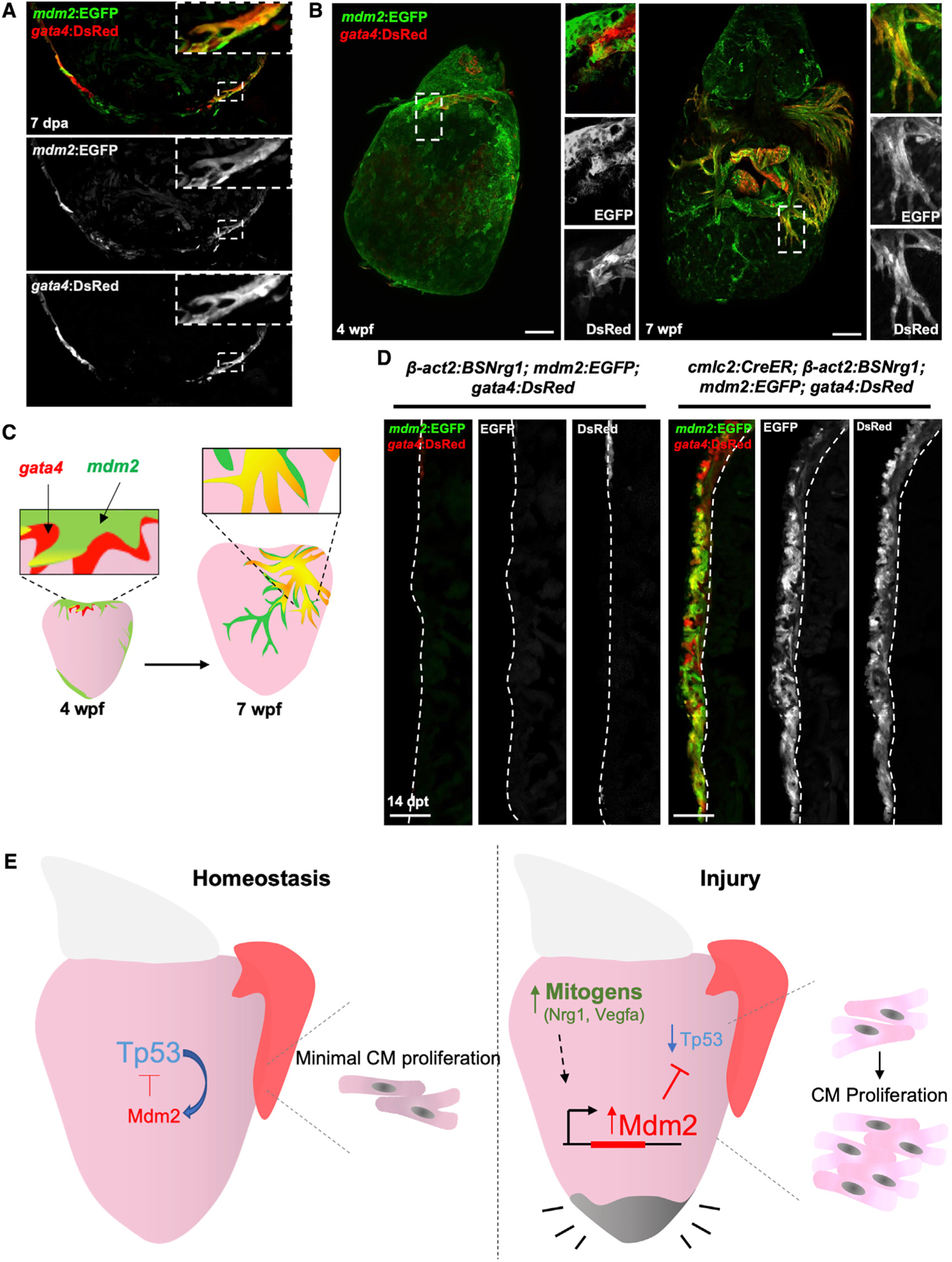
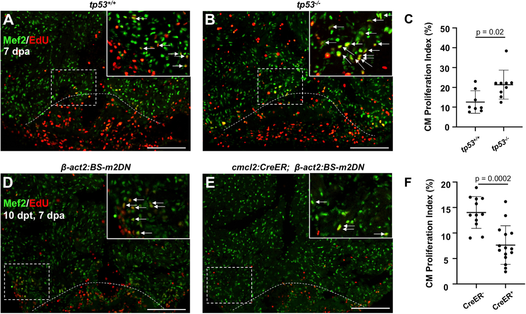
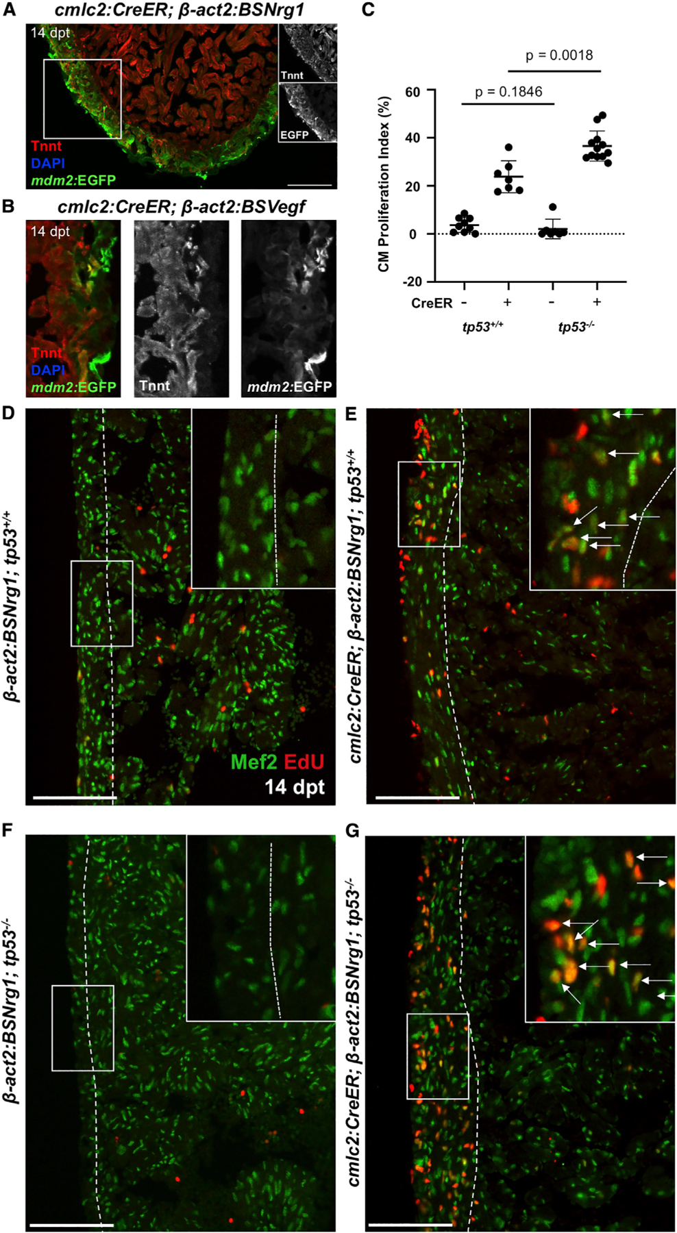
Zebrafish regenerate heart muscle through division of pre-existing cardiomyocytes. To discover underlying regulation, we assess transcriptome datasets for dynamic gene networks during heart regeneration and identify suppression of genes associated with the transcription factor Tp53. Cardiac damage leads to fluctuation of Tp53 protein levels, concomitant with induced expression of its central negative regulator, mdm2, in regenerating cardiomyocytes. Zebrafish lacking functional Tp53 display increased indicators of cardiomyocyte proliferation during regeneration, whereas transgenic Mdm2 blockade inhibits injury-induced cardiomyocyte proliferation. Induced myocardial overexpression of the mitogenic factors Nrg1 or Vegfaa in the absence of injury also upregulates mdm2 and suppresses Tp53 levels, and tp53 mutations augment the mitogenic effects of Nrg1. mdm2 induction is spatiotemporally associated with markers of de-differentiation in injury and growth contexts, suggesting a broad role in cardiogenesis. Our findings reveal myocardial Tp53 suppression by mitogen-induced Mdm2 as a regulatory component of innate cardiac regeneration.

摘要

斑马鱼通过已有心肌细胞的分裂来再生心肌。为了发现潜在的调控机制,我们评估了心脏再生过程中动态基因网络的转录组数据集,并鉴定出与转录因子 Tp53 相关的基因受到抑制。心脏损伤导致 Tp53 蛋白水平波动,同时在再生心肌细胞中诱导其核心负调控因子 mdm2 的表达。缺乏功能性 Tp53 的斑马鱼在再生过程中显示出心肌细胞增殖的增加指标,而转基因 Mdm2 阻断则抑制损伤诱导的心肌细胞增殖。在没有损伤的情况下,诱导心肌中促有丝分裂因子 Nrg1 或 Vegfaa 的过度表达也会上调 mdm2 并抑制 Tp53 水平,而 tp53 突变会增强 Nrg1 的促有丝分裂作用。mdm2 的诱导与损伤和生长环境中去分化标记物在时空上相关,提示其在心脏发生中具有广泛的作用。我们的研究结果揭示了心肌细胞中由有丝分裂原诱导的 Mdm2 对 Tp53 的抑制作用,这是心脏再生内在调节的一个组成部分。

https://cdn.ncbi.nlm.nih.gov/pmc/blobs/ee58/7494019/1731563c680b/nihms-1627845-f0005.jpg
https://cdn.ncbi.nlm.nih.gov/pmc/blobs/ee58/7494019/ee1297987783/nihms-1627845-f0002.jpg
https://cdn.ncbi.nlm.nih.gov/pmc/blobs/ee58/7494019/92ac50cfe61c/nihms-1627845-f0003.jpg
https://cdn.ncbi.nlm.nih.gov/pmc/blobs/ee58/7494019/12540e72ded8/nihms-1627845-f0004.jpg
https://cdn.ncbi.nlm.nih.gov/pmc/blobs/ee58/7494019/1731563c680b/nihms-1627845-f0005.jpg

相似文献

1
Tp53 Suppression Promotes Cardiomyocyte Proliferation during Zebrafish Heart Regeneration.抑癌基因 TP53 促进斑马鱼心脏再生过程中心肌细胞的增殖。
Cell Rep. 2020 Sep 1;32(9):108089. doi: 10.1016/j.celrep.2020.108089.
2
Nrg1 is an injury-induced cardiomyocyte mitogen for the endogenous heart regeneration program in zebrafish.神经调节蛋白1(Nrg1)是一种损伤诱导的心肌细胞有丝分裂原,用于斑马鱼体内的心脏再生程序。
Elife. 2015 Apr 1;4:e05871. doi: 10.7554/eLife.05871.
3
Primary contribution to zebrafish heart regeneration by gata4(+) cardiomyocytes.Gata4(+) 心肌细胞对斑马鱼心脏再生的主要贡献。
Nature. 2010 Mar 25;464(7288):601-5. doi: 10.1038/nature08804.
4
Single-cell analysis uncovers that metabolic reprogramming by ErbB2 signaling is essential for cardiomyocyte proliferation in the regenerating heart.单细胞分析揭示了 ErbB2 信号转导的代谢重编程对于心脏再生过程中心肌细胞的增殖是必不可少的。
Elife. 2019 Dec 23;8:e50163. doi: 10.7554/eLife.50163.
5
Fibronectin is deposited by injury-activated epicardial cells and is necessary for zebrafish heart regeneration.纤维连接蛋白由损伤激活的心脏外膜细胞分泌,并对斑马鱼心脏再生是必需的。
Dev Biol. 2013 Oct 15;382(2):427-35. doi: 10.1016/j.ydbio.2013.08.012. Epub 2013 Aug 26.
6
Cardiomyocyte heterogeneity during zebrafish development and regeneration.斑马鱼发育和再生过程中心肌细胞的异质性。
Dev Biol. 2021 Aug;476:259-271. doi: 10.1016/j.ydbio.2021.03.014. Epub 2021 Apr 12.
7
Vegfaa instructs cardiac muscle hyperplasia in adult zebrafish.Vegfaa 指导成年斑马鱼心肌细胞增生。
Proc Natl Acad Sci U S A. 2018 Aug 28;115(35):8805-8810. doi: 10.1073/pnas.1722594115. Epub 2018 Aug 13.
8
In vivo proximity labeling identifies cardiomyocyte protein networks during zebrafish heart regeneration.体内邻近标记法可识别斑马鱼心脏再生过程中的心肌细胞蛋白质网络。
Elife. 2021 Mar 25;10:e66079. doi: 10.7554/eLife.66079.
9
Igf Signaling is Required for Cardiomyocyte Proliferation during Zebrafish Heart Development and Regeneration.胰岛素样生长因子信号传导在斑马鱼心脏发育和再生过程中对心肌细胞增殖是必需的。
PLoS One. 2013 Jun 26;8(6):e67266. doi: 10.1371/journal.pone.0067266. Print 2013.
10
Hand2 elevates cardiomyocyte production during zebrafish heart development and regeneration.HAND2 促进斑马鱼心脏发育和再生过程中心肌细胞的生成。
Development. 2014 Aug;141(16):3112-22. doi: 10.1242/dev.106336. Epub 2014 Jul 18.

引用本文的文献

1
Newborn apical resection preserves the proliferative capacity of cardiomyocytes located throughout the left ventricle.新生儿心尖切除术可保留整个左心室内心肌细胞的增殖能力。
Stem Cells. 2025 May 15;43(5). doi: 10.1093/stmcls/sxaf018.
2
Competitive signaling and cellular communications in myocardial infarction response.心肌梗死反应中的竞争性信号传导与细胞通讯
Mol Biol Rep. 2025 Jan 16;52(1):129. doi: 10.1007/s11033-025-10236-5.
3
Roles of naïve CD4 T cells and their differentiated subtypes in lung adenocarcinoma and underlying potential regulatory pathways.
幼稚 CD4 T 细胞及其分化亚型在肺腺癌中的作用及潜在的调控途径。
J Transl Med. 2024 Aug 22;22(1):781. doi: 10.1186/s12967-024-05530-y.
4
P53 Activation Promotes Maturational Characteristics of Pluripotent Stem Cell-Derived Cardiomyocytes in 3-Dimensional Suspension Culture Via FOXO-FOXM1 Regulation.P53 激活通过 FOXO-FOXM1 调控促进三维悬浮培养多能干细胞源性心肌细胞的成熟特征。
J Am Heart Assoc. 2024 Jul 2;13(13):e033155. doi: 10.1161/JAHA.123.033155. Epub 2024 Jun 27.
5
Comparative Analysis of Heart Regeneration: Searching for the Key to Heal the Heart-Part II: Molecular Mechanisms of Cardiac Regeneration.心脏再生的比较分析:探寻治愈心脏的关键——第二部分:心脏再生的分子机制
J Cardiovasc Dev Dis. 2023 Aug 22;10(9):357. doi: 10.3390/jcdd10090357.
6
Spinal cord repair is modulated by the neurogenic factor Hb-egf under direction of a regeneration-associated enhancer.脊髓修复受神经发生因子 Hb-egf 调节,受与再生相关增强子的指导。
Nat Commun. 2023 Aug 11;14(1):4857. doi: 10.1038/s41467-023-40486-5.
7
Cardiomyocyte Ploidy, Metabolic Reprogramming and Heart Repair.心肌细胞倍性、代谢重编程与心脏修复。
Cells. 2023 Jun 7;12(12):1571. doi: 10.3390/cells12121571.
8
Polycomb Group Protein CBX7 Represses Cardiomyocyte Proliferation Through Modulation of the TARDBP/RBM38 Axis.多梳蛋白 CBX7 通过调节 TARDBP/RBM38 轴抑制心肌细胞增殖。
Circulation. 2023 Jun 13;147(24):1823-1842. doi: 10.1161/CIRCULATIONAHA.122.061131. Epub 2023 May 9.
9
Transcriptomic data meta-analysis reveals common and injury model specific gene expression changes in the regenerating zebrafish heart.转录组数据分析揭示了再生斑马鱼心脏中共同的和损伤模型特异的基因表达变化。
Sci Rep. 2023 Apr 3;13(1):5418. doi: 10.1038/s41598-023-32272-6.
10
Exploring the Potential Mechanism of Qi-Shen-Di-Huang Drug Formulary for Myasthenia Gravis (MG) based on UHPLC-QE-MS Network Pharmacology and Molecular Docking Techniques.基于超高效液相色谱-四极杆飞行时间质谱联用网络药理学和分子对接技术探索杞参地黄方治疗重症肌无力的潜在机制
Evid Based Complement Alternat Med. 2022 Oct 3;2022:7416448. doi: 10.1155/2022/7416448. eCollection 2022.