• 文献检索
  • 文档翻译
  • 深度研究
  • 学术资讯
  • Suppr Zotero 插件Zotero 插件
  • 邀请有礼
  • 套餐&价格
  • 历史记录
应用&插件
Suppr Zotero 插件Zotero 插件浏览器插件Mac 客户端Windows 客户端微信小程序
定价
高级版会员购买积分包购买API积分包
服务
文献检索文档翻译深度研究API 文档MCP 服务
关于我们
关于 Suppr公司介绍联系我们用户协议隐私条款
关注我们

Suppr 超能文献

核心技术专利:CN118964589B侵权必究
粤ICP备2023148730 号-1Suppr @ 2026

文献检索

告别复杂PubMed语法,用中文像聊天一样搜索,搜遍4000万医学文献。AI智能推荐,让科研检索更轻松。

立即免费搜索

文件翻译

保留排版,准确专业,支持PDF/Word/PPT等文件格式,支持 12+语言互译。

免费翻译文档

深度研究

AI帮你快速写综述,25分钟生成高质量综述,智能提取关键信息,辅助科研写作。

立即免费体验

Notch4 通过 ERK/JNK/P38MAPK 信号通路介导低氧性肺动脉高压中的血管重塑。

Notch4 mediates vascular remodeling via ERK/JNK/P38 MAPK signaling pathways in hypoxic pulmonary hypertension.

机构信息

Department of Pulmonary and Critical Care Medicine, Tongji Hospital, Tongji Medical College, Huazhong University of Science and Technology, 1095 Jiefang Avenue, Wuhan, 430030, China.

Key Laboratory of Respiratory Diseases, National Ministry of Health of the People's Republic of China and National Clinical Research Center for Respiratory Disease, Wuhan, China.

出版信息

Respir Res. 2022 Jan 11;23(1):6. doi: 10.1186/s12931-022-01927-9.

DOI:10.1186/s12931-022-01927-9
PMID:35016680
原文链接:https://pmc.ncbi.nlm.nih.gov/articles/PMC8753901/
Abstract

BACKGROUND

Hypoxic pulmonary hypertension (HPH) is a chronic progressive advanced disorder pathologically characterized by pulmonary vascular remodeling. Notch4 as a cell surface receptor is critical for vascular development. However, little is known about the role and mechanism of Notch4 in the development of hypoxic vascular remodeling.

METHODS

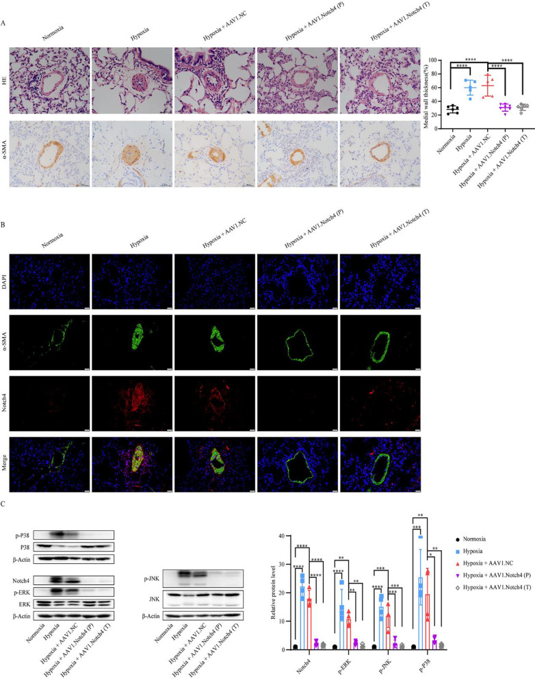
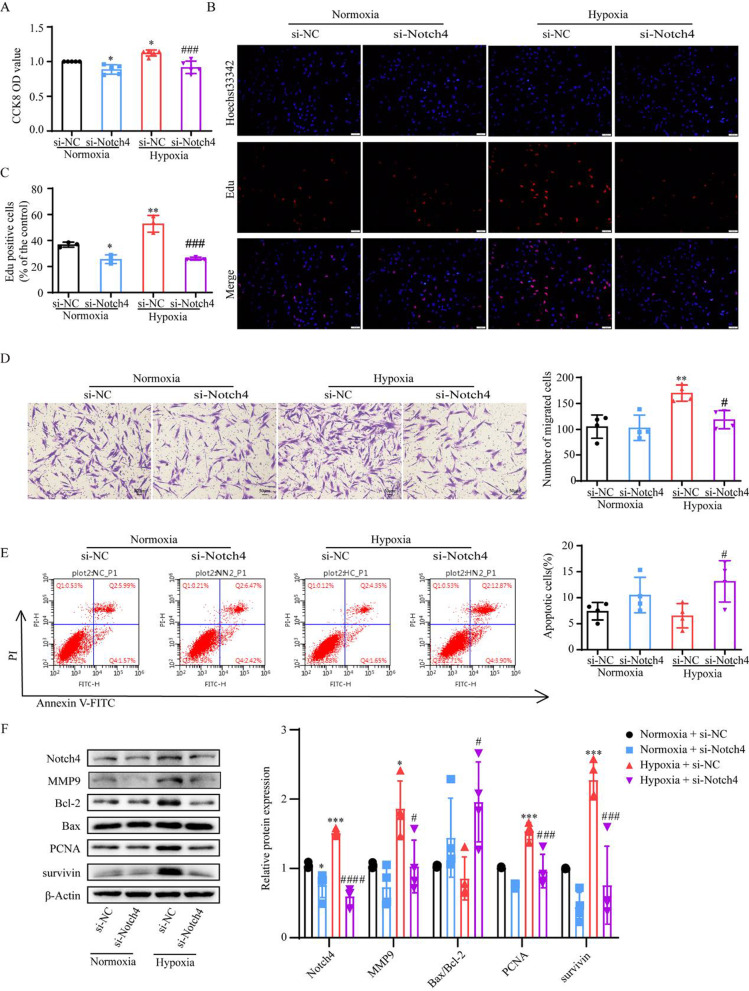
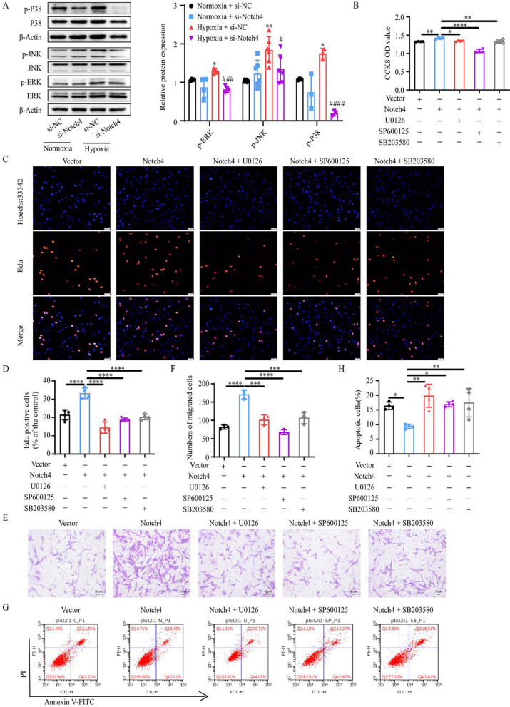
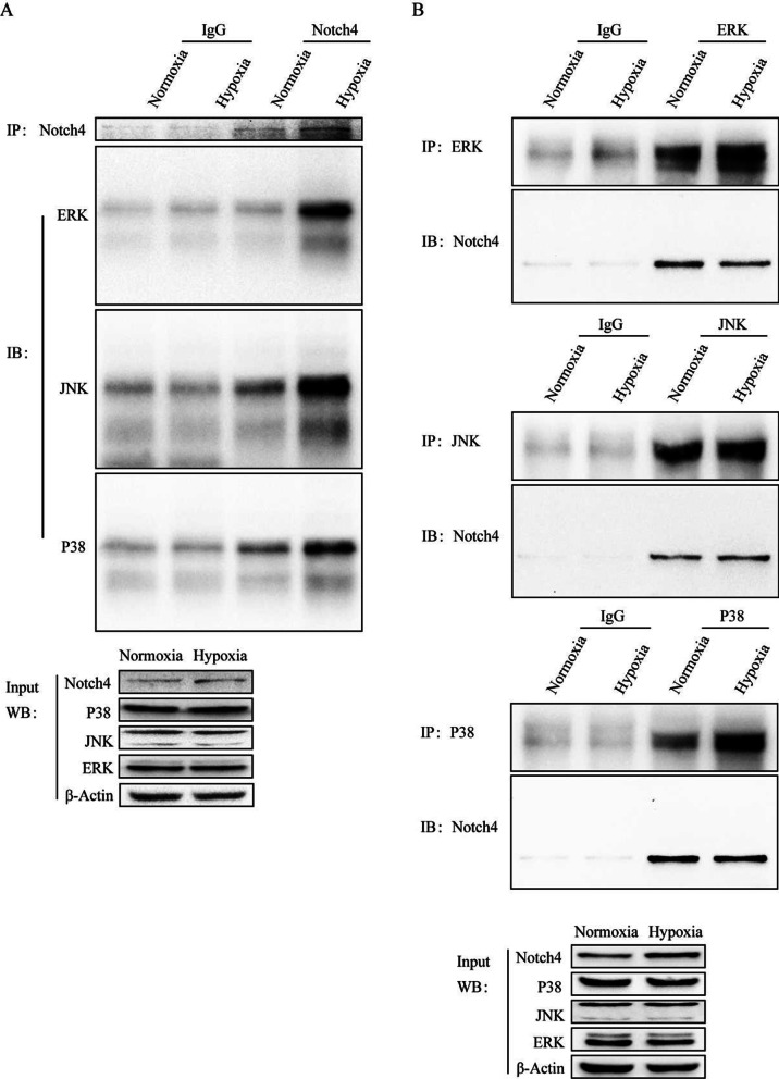
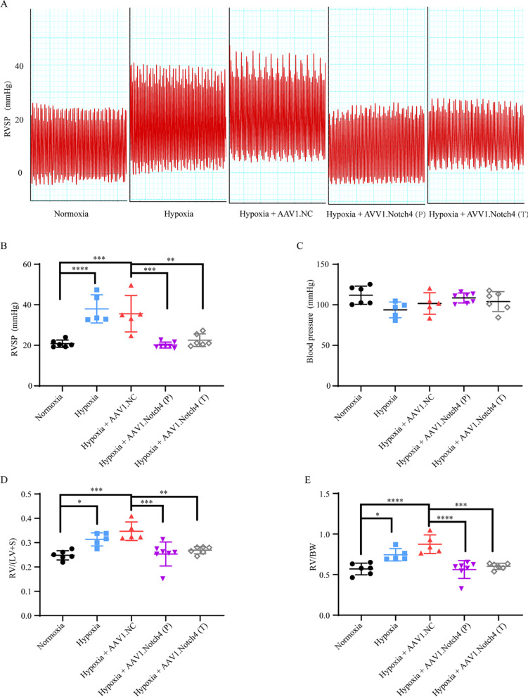
Lung tissue samples were collected to detect the expression of Notch4 from patients with HPH and matched controls. Human pulmonary artery smooth muscle cells (HPASMCs) were cultured in hypoxic and normoxic conditions. Real-time quantitative PCR and western blotting were used to examine the mRNA and protein levels of Notch4. HPASMCs were transfected with small interference RNA (siRNA) against Notch4 or Notch4 overexpression plasmid, respectively. Cell viability, cell proliferation, apoptosis, and migration were assessed using Cell Counting Kit-8, Edu, Annexin-V/PI, and Transwell assay. The interaction between Notch4 and ERK, JNK, P38 MAPK were analyzed by co-immunoprecipitation. Adeno-associated virus 1-mediated siRNA against Notch4 (AAV1-si-Notch4) was injected into the airways of hypoxic rats. Right ventricular systolic pressure (RVSP), right ventricular hypertrophy and pulmonary vascular remodeling were evaluated.

RESULTS

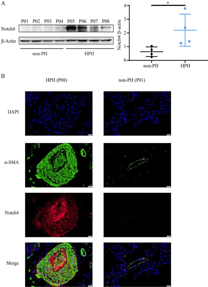
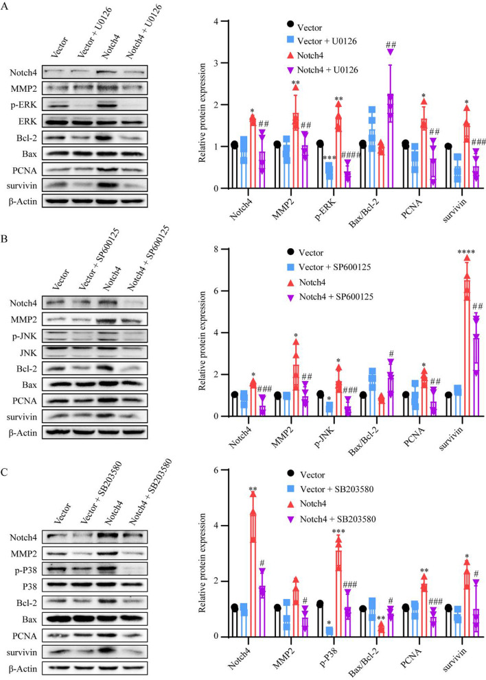
In this study, we demonstrate that Notch4 is highly expressed in the media of pulmonary vascular and is upregulated in lung tissues from patients with HPH and HPH rats compared with control groups. In vitro, hypoxia induces the high expression of Delta-4 and Notch4 in HPASMCs. The increased expression of Notch4 promotes HPASMCs proliferation and migration and inhibits cells apoptosis via ERK, JNK, P38 signaling pathways. Furthermore, co-immunoprecipitation result elucidates the interaction between Notch4 and ERK/JNK/P38. In vivo, silencing Notch4 partly abolished the increase in RVSP and pulmonary vascular remodeling caused by hypoxia in HPH rats.

CONCLUSIONS

These findings reveal an important role of the Notch4-ERK/JNK/P38 MAPK axis in hypoxic pulmonary remodeling and provide a potential therapeutic target for patients with HPH.

摘要

背景

低氧性肺动脉高压(HPH)是一种慢性进行性进展性疾病,其病理特征为肺血管重构。Notch4 作为细胞表面受体,对血管发育至关重要。然而,Notch4 在低氧性血管重构发展中的作用和机制知之甚少。

方法

收集 HPH 患者和匹配对照的肺组织样本,检测 Notch4 的表达。在低氧和常氧条件下培养人肺动脉平滑肌细胞(HPASMCs)。实时定量 PCR 和 Western blot 检测 Notch4 的 mRNA 和蛋白水平。分别用 Notch4 小干扰 RNA(siRNA)或 Notch4 过表达质粒转染 HPASMCs。用细胞计数试剂盒-8(Cell Counting Kit-8)、Edu、Annexin-V/PI 和 Transwell 检测细胞活力、增殖、凋亡和迁移。通过共免疫沉淀分析 Notch4 与 ERK、JNK、P38 MAPK 的相互作用。将腺相关病毒 1 介导的 Notch4 siRNA(AAV1-si-Notch4)注入低氧大鼠气道。评估右心室收缩压(RVSP)、右心室肥厚和肺血管重构。

结果

本研究表明,Notch4 在肺血管中层高度表达,并在 HPH 患者和 HPH 大鼠的肺组织中上调,高于对照组。体外,低氧诱导 HPASMCs 中 Delta-4 和 Notch4 的高表达。Notch4 的高表达促进 HPASMCs 增殖和迁移,通过 ERK、JNK、P38 信号通路抑制细胞凋亡。此外,共免疫沉淀结果阐明了 Notch4 与 ERK/JNK/P38 的相互作用。在体内,沉默 Notch4 部分阻断了 HPH 大鼠低氧引起的 RVSP 升高和肺血管重构。

结论

这些发现揭示了 Notch4-ERK/JNK/P38 MAPK 轴在低氧性肺重构中的重要作用,为 HPH 患者提供了一个潜在的治疗靶点。

https://cdn.ncbi.nlm.nih.gov/pmc/blobs/e21d/8753901/186606bbdc9d/12931_2022_1927_Fig7_HTML.jpg
https://cdn.ncbi.nlm.nih.gov/pmc/blobs/e21d/8753901/062824478ef2/12931_2022_1927_Fig1_HTML.jpg
https://cdn.ncbi.nlm.nih.gov/pmc/blobs/e21d/8753901/b7d2b54537c3/12931_2022_1927_Fig2_HTML.jpg
https://cdn.ncbi.nlm.nih.gov/pmc/blobs/e21d/8753901/1143b3ef5901/12931_2022_1927_Fig3_HTML.jpg
https://cdn.ncbi.nlm.nih.gov/pmc/blobs/e21d/8753901/c0b50c9cf487/12931_2022_1927_Fig4_HTML.jpg
https://cdn.ncbi.nlm.nih.gov/pmc/blobs/e21d/8753901/36dffa2439e5/12931_2022_1927_Fig5_HTML.jpg
https://cdn.ncbi.nlm.nih.gov/pmc/blobs/e21d/8753901/f3865b9b4957/12931_2022_1927_Fig6_HTML.jpg
https://cdn.ncbi.nlm.nih.gov/pmc/blobs/e21d/8753901/186606bbdc9d/12931_2022_1927_Fig7_HTML.jpg

相似文献

1
Notch4 mediates vascular remodeling via ERK/JNK/P38 MAPK signaling pathways in hypoxic pulmonary hypertension.Notch4 通过 ERK/JNK/P38MAPK 信号通路介导低氧性肺动脉高压中的血管重塑。
Respir Res. 2022 Jan 11;23(1):6. doi: 10.1186/s12931-022-01927-9.
2
TRB3 mediates vascular remodeling by activating the MAPK signaling pathway in hypoxic pulmonary hypertension.TRB3 通过激活低氧性肺动脉高压中的 MAPK 信号通路来介导血管重构。
Respir Res. 2021 Dec 14;22(1):312. doi: 10.1186/s12931-021-01908-4.
3
MiR-593-5p promotes the development of hypoxic-induced pulmonary hypertension via targeting PLK1.miR-593-5p 通过靶向 PLK1 促进低氧诱导的肺动脉高压的发展。
Eur Rev Med Pharmacol Sci. 2019 Apr;23(8):3495-3502. doi: 10.26355/eurrev_201904_17715.
4
The effect of activated κ-opioid receptor (κ-OR) on the role of calcium sensing receptor (CaSR) in preventing hypoxic pulmonary hypertension development.激活 κ-阿片受体(κ-OR)对钙敏感受体(CaSR)在预防低氧性肺动脉高压发展中的作用的影响。
Biomed Pharmacother. 2020 May;125:109931. doi: 10.1016/j.biopha.2020.109931. Epub 2020 Feb 14.
5
Exosomes derived from mesenchymal stromal cells exert a therapeutic effect on hypoxia-induced pulmonary hypertension by modulating the YAP1/SPP1 signaling pathway.间充质基质细胞衍生的外泌体通过调节 YAP1/SPP1 信号通路对低氧诱导的肺动脉高压发挥治疗作用。
Biomed Pharmacother. 2023 Dec;168:115816. doi: 10.1016/j.biopha.2023.115816. Epub 2023 Nov 2.
6
Proteomic analysis reveals that Xbp1s promotes hypoxic pulmonary hypertension through the p-JNK MAPK pathway.蛋白质组学分析表明,Xbp1s通过p-JNK丝裂原活化蛋白激酶途径促进缺氧性肺动脉高压。
J Cell Physiol. 2022 Mar;237(3):1948-1963. doi: 10.1002/jcp.30664. Epub 2021 Dec 28.
7
The alveolar epithelial cells are involved in pulmonary vascular remodeling and constriction of hypoxic pulmonary hypertension.肺泡上皮细胞参与肺部血管重构和低氧性肺动脉高压的收缩。
Respir Res. 2021 May 4;22(1):134. doi: 10.1186/s12931-021-01708-w.
8
Involvement of Gax gene in hypoxia-induced pulmonary hypertension, proliferation, and apoptosis of arterial smooth muscle cells.Gax 基因在低氧诱导的肺动脉高压、动脉平滑肌细胞增殖和凋亡中的作用。
Am J Respir Cell Mol Biol. 2011 Jan;44(1):66-73. doi: 10.1165/rcmb.2008-0442OC. Epub 2010 Feb 16.
9
NDUFA4L2 in smooth muscle promotes vascular remodeling in hypoxic pulmonary arterial hypertension.NDUFA4L2 在平滑肌中促进低氧性肺动脉高压中的血管重构。
J Cell Mol Med. 2021 Jan;25(2):1221-1237. doi: 10.1111/jcmm.16193. Epub 2020 Dec 19.
10
Nur77 induced by HIF-1α mediates vascular remodeling in hypoxic pulmonary hypertension.缺氧诱导因子-1α诱导 Nur77 介导线粒体凋亡参与低氧性肺动脉高压血管重构。
Cell Mol Biol (Noisy-le-grand). 2024 Jun 5;70(6):233-237. doi: 10.14715/cmb/2024.70.6.35.

引用本文的文献

1
Hypoxia- and mechanical stress-induced upregulation of mitochondrial HSP60 is associated with phenotypic switching of pulmonary arterial smooth muscle cells.缺氧和机械应激诱导的线粒体热休克蛋白60上调与肺动脉平滑肌细胞的表型转换有关。
Cell Stress Chaperones. 2025 Jul;30(4):100089. doi: 10.1016/j.cstres.2025.100089. Epub 2025 Jul 2.
2
Signaling pathways and targeted therapy for pulmonary hypertension.肺动脉高压的信号通路与靶向治疗
Signal Transduct Target Ther. 2025 Jul 1;10(1):207. doi: 10.1038/s41392-025-02287-8.
3
Bronchial anastomotic complications as a microvascular disruption in a mouse model of airway transplantation.

本文引用的文献

1
Targeting oncogenic Notch signaling with SERCA inhibitors.针对致癌性 Notch 信号通路的 SERCA 抑制剂。
J Hematol Oncol. 2021 Jan 6;14(1):8. doi: 10.1186/s13045-020-01015-9.
2
Resistin-like molecule β acts as a mitogenic factor in hypoxic pulmonary hypertension via the Ca-dependent PI3K/Akt/mTOR and PKC/MAPK signaling pathways.抵抗素样分子 β 通过 Ca 依赖性 PI3K/Akt/mTOR 和 PKC/MAPK 信号通路在低氧性肺动脉高压中作为有丝分裂因子发挥作用。
Respir Res. 2021 Jan 6;22(1):8. doi: 10.1186/s12931-020-01598-4.
3
The preventive and therapeutic effects of AAV1-KLF4-shRNA in cigarette smoke-induced pulmonary hypertension.
支气管吻合口并发症作为气道移植小鼠模型中的微血管破坏。
Front Immunol. 2025 May 14;16:1567657. doi: 10.3389/fimmu.2025.1567657. eCollection 2025.
4
Silencing PPAP2C inhibits lung adenocarcinoma migration and invasion via the ERK/JNK pathway.沉默 PPAP2C 通过 ERK/JNK 通路抑制肺腺癌迁移和侵袭。
Mol Med Rep. 2025 Jan;31(1). doi: 10.3892/mmr.2024.13392. Epub 2024 Nov 14.
5
Early short-term hypoxia promotes epidermal cell migration by activating the CCL2-ERK1/2 pathway and epithelial-mesenchymal transition during wound healing.早期短期缺氧通过激活CCL2-ERK1/2通路和上皮-间质转化促进伤口愈合过程中的表皮细胞迁移。
Burns Trauma. 2024 Jun 17;12:tkae017. doi: 10.1093/burnst/tkae017. eCollection 2024.
6
Differential expression and analysis of extrachromosomal circular DNAs as serum biomarkers in pulmonary arterial hypertension.肺动脉高压血清生物标志物中外环 DNA 的差异表达与分析。
Respir Res. 2024 Apr 25;25(1):181. doi: 10.1186/s12931-024-02808-z.
7
Up-regulated expression of two-pore domain K channels, KCNK1 and KCNK2, is involved in the proliferation and migration of pulmonary arterial smooth muscle cells in pulmonary arterial hypertension.双孔域钾通道KCNK1和KCNK2的表达上调参与肺动脉高压中肺动脉平滑肌细胞的增殖和迁移。
Front Cardiovasc Med. 2024 Feb 12;11:1343804. doi: 10.3389/fcvm.2024.1343804. eCollection 2024.
8
Ginkgolide B Blocks Vascular Remodeling after Vascular Injury via Regulating Tgf1/Smad Signaling Pathway.银杏内酯 B 通过调控 TGF1/Smad 信号通路抑制血管损伤后血管重构。
Cardiovasc Ther. 2023 Dec 13;2023:8848808. doi: 10.1155/2023/8848808. eCollection 2023.
9
CC chemokines Modulate Immune responses in Pulmonary Hypertension.CC 趋化因子调节肺动脉高压中的免疫反应。
J Adv Res. 2024 Sep;63:171-186. doi: 10.1016/j.jare.2023.10.015. Epub 2023 Nov 4.
10
Insights into Endothelin Receptors in Pulmonary Hypertension.肺动脉高压中内皮素受体的研究进展。
Int J Mol Sci. 2023 Jun 16;24(12):10206. doi: 10.3390/ijms241210206.
AAV1-KLF4-shRNA 在香烟烟雾诱导的肺动脉高压中的预防和治疗作用。
J Cell Mol Med. 2021 Jan;25(2):1238-1251. doi: 10.1111/jcmm.16194. Epub 2020 Dec 20.
4
Regulation of circular RNAs act as ceRNA in a hypoxic pulmonary hypertension rat model.环状 RNA 作为 ceRNA 在低氧性肺动脉高压大鼠模型中的作用。
Genomics. 2021 Jan;113(1 Pt 1):11-19. doi: 10.1016/j.ygeno.2020.11.021. Epub 2020 Nov 27.
5
Notch3 signalling and vascular remodelling in pulmonary arterial hypertension.Notch3 信号通路与肺动脉高压中的血管重构。
Clin Sci (Lond). 2019 Dec 20;133(24):2481-2498. doi: 10.1042/CS20190835.
6
Overlapping but distinct roles for NOTCH receptors in human cardiovascular disease.NOTCH 受体在人类心血管疾病中具有重叠但不同的作用。
Clin Genet. 2019 Jan;95(1):85-94. doi: 10.1111/cge.13382. Epub 2018 Jun 10.
7
Overcoming the Host Immune Response to Adeno-Associated Virus Gene Delivery Vectors: The Race Between Clearance, Tolerance, Neutralization, and Escape.克服腺相关病毒基因传递载体的宿主免疫反应:清除、耐受、中和和逃逸之间的竞赛。
Annu Rev Virol. 2017 Sep 29;4(1):511-534. doi: 10.1146/annurev-virology-101416-041936.
8
Unraveling the Complex Story of Immune Responses to AAV Vectors Trial After Trial.一项又一项试验揭示腺相关病毒载体免疫反应的复杂故事
Hum Gene Ther. 2017 Nov;28(11):1061-1074. doi: 10.1089/hum.2017.150.
9
Notch-4 silencing inhibits prostate cancer growth and EMT via the NF-κB pathway.Notch-4基因沉默通过NF-κB信号通路抑制前列腺癌生长和上皮间质转化。
Apoptosis. 2017 Jun;22(6):877-884. doi: 10.1007/s10495-017-1368-0.
10
Pulmonary Hypertension.肺动脉高压。
Dtsch Arztebl Int. 2017 Feb 3;114(5):73-84. doi: 10.3238/arztebl.2017.0073.