Department of Traumatology, Sestre milosrdnice University Hospital Center, Draškovićeva 19, 10000, Zagreb, Croatia.
University of Applied Health Sciences, Mlinarska cesta 38, 10000, Zagreb, Croatia.
J Orthop Surg Res. 2022 Jan 11;17(1):16. doi: 10.1186/s13018-021-02907-3.
Good clinical outcomes for locking plates as an external fixator to treat tibial fractures have been reported. However, external locking plate fixation is still generally rarely performed. This study aimed to compare the stability of an external locking plate fixator with that of a conventional external fixator for extraarticular proximal tibial fractures using finite element analysis.
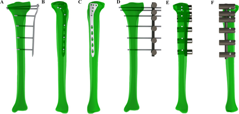
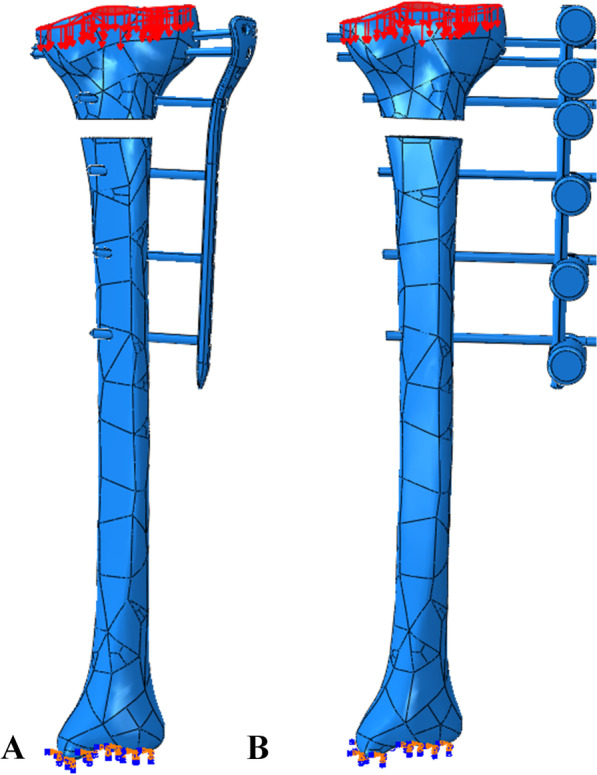
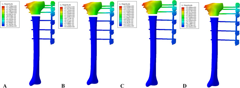
Three models were constructed: (1) external locking plate fixation of proximal tibial fracture with lateral proximal tibial locking plate and 5-mm screws (ELP), (2) conventional external fixation of proximal tibial fracture with an 11-mm rod and 5-mm Schanz screws (EF-11), and (3) conventional external fixation of a proximal tibial fracture with a 7-mm rod and 5-mm Schanz screws (EF-7). The stress distribution, displacement at the fracture gap, and stiffness of the three finite element models at 30-, 40-, 50-, and 60-mm plate-rod offsets from the lateral surface of the lateral condyle of the tibia were determined.
The conventional external fixator showed higher stiffness than the external locking plate fixator. In all models, the stiffness decreased as the distance of the plate-rod from the bone surface increased. The maximum stiffness was 121.06 N/mm in the EF-11 model with 30-mm tibia-rod offset. In the EF-7 model group, the maximum stiffness was 40.00 N/mm in the model with 30-mm tibia-rod offset. In the ELP model group, the maximum stiffness was 35.79 N/mm in the model with 30-mm tibia-plate offset.
Finite element analysis indicated that external locking plate fixation is more flexible than conventional external fixation and can influence secondary bone healing. External locking plate fixation requires the placement of the plate as close as possible to the skin, which allows for a low-profile design because the increased distance from the plate to the bone can be too flexible for bone healing. Further experimental mechanical model tests are necessary to validate these finite element models, and further biological analysis is necessary to evaluate the effect of external locking plate fixation on fracture healing.
已有研究报道锁定钢板作为外固定器治疗胫骨骨折具有良好的临床效果。然而,外锁钢板固定术通常仍很少应用。本研究旨在通过有限元分析比较外锁钢板固定器与传统外固定器治疗胫骨关节外近端骨折的稳定性。
构建了 3 种模型:(1)胫骨外侧近端锁定钢板和 5mm 螺钉的胫骨近端骨折外锁钢板固定(ELP);(2)11mm 杆和 5mm Schanz 螺钉的胫骨近端骨折传统外固定(EF-11);(3)7mm 杆和 5mm Schanz 螺钉的胫骨近端骨折传统外固定(EF-7)。测定了 3 种有限元模型在胫骨外侧髁外侧 30、40、50、60mm 处的骨折间隙位移和板-杆偏移的骨折间隙的应力分布和刚度。
传统外固定器的刚度高于外锁钢板固定器。在所有模型中,随着板-杆距骨表面距离的增加,刚度减小。EF-11 模型中,胫骨-杆偏移 30mm 时的最大刚度为 121.06N/mm。在 EF-7 模型组中,胫骨-杆偏移 30mm 时的最大刚度为 40.00N/mm。在 ELP 模型组中,胫骨-板偏移 30mm 时的最大刚度为 35.79N/mm。
有限元分析表明,外锁钢板固定比传统外固定更具灵活性,并可能影响二次骨愈合。外锁钢板固定要求将钢板尽可能靠近皮肤放置,这允许采用低轮廓设计,因为钢板与骨之间的距离增加会使骨愈合过于灵活。需要进一步进行实验力学模型试验来验证这些有限元模型,并进一步进行生物分析来评估外锁钢板固定对骨折愈合的影响。