• 文献检索
  • 文档翻译
  • 深度研究
  • 学术资讯
  • Suppr Zotero 插件Zotero 插件
  • 邀请有礼
  • 套餐&价格
  • 历史记录
应用&插件
Suppr Zotero 插件Zotero 插件浏览器插件Mac 客户端Windows 客户端微信小程序
定价
高级版会员购买积分包购买API积分包
服务
文献检索文档翻译深度研究API 文档MCP 服务
关于我们
关于 Suppr公司介绍联系我们用户协议隐私条款
关注我们

Suppr 超能文献

核心技术专利:CN118964589B侵权必究
粤ICP备2023148730 号-1Suppr @ 2026

文献检索

告别复杂PubMed语法,用中文像聊天一样搜索,搜遍4000万医学文献。AI智能推荐,让科研检索更轻松。

立即免费搜索

文件翻译

保留排版,准确专业,支持PDF/Word/PPT等文件格式,支持 12+语言互译。

免费翻译文档

深度研究

AI帮你快速写综述,25分钟生成高质量综述,智能提取关键信息,辅助科研写作。

立即免费体验

脑出血中的缺血:小血管和大血管疾病的比较研究。

Ischemia in intracerebral hemorrhage: A comparative study of small-vessel and large-vessel diseases.

机构信息

Department of Neurology, People's Hospital of Zhengzhou, Zhengzhou, China.

Department of Neurology, People's Hospital of Henan University of Chinese Medicine, Zhengzhou, China.

出版信息

Ann Clin Transl Neurol. 2022 Jan;9(1):79-90. doi: 10.1002/acn3.51497. Epub 2022 Jan 12.

DOI:10.1002/acn3.51497
PMID:35018741
原文链接:https://pmc.ncbi.nlm.nih.gov/articles/PMC8791802/
Abstract

OBJECTIVE

This study aimed to compare effects of cerebral small-vessel disease (cSVD) burden and cerebral artery stenosis (CAS) on acute ischemia in intracerebral hemorrhage (ICH) and their interaction with mean arterial pressure (MAP) change.

METHODS

We recruited consecutive patients with acute primary ICH. Brain magnetic resonance imaging and angiography were performed to quantify diffusion-weighted imaging (DWI) lesions, CAS, and cSVD markers, which were calculated for the total cSVD score. Multivariable regression models were adopted to explore their associations by DWI lesions size (<15 vs. ≥15 mm) and median MAP change stratification.

RESULTS

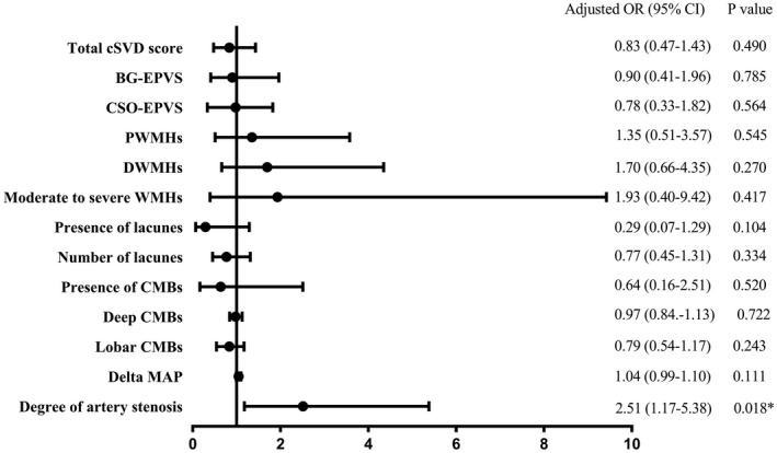
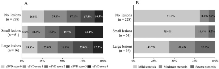
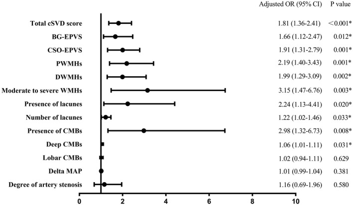
Of 305 included patients (mean age 59.5 years, 67.9% males), 77 (25.2%) had DWI lesions (small, 79.2%; large, 20.8%) and 67 (22.0%) had moderate and severe CAS. In multivariable analysis, small DWI lesions were independently associated with higher total cSVD score (odds ratio [OR] 1.81, 95% confidence interval [CI] 1.36-2.41). and large DWI lesions were associated with more severe CAS (OR 2.51, 95% CI 1.17-5.38). This association was modified by MAP change (interaction p = 0.016), with stratified analysis showing an increased risk of large DWI lesions in severe CAS with greater MAP change (≥44 mmHg) (OR 3.48, 95% CI 1.13-10.74) but not with mild MAP change (<44 mmHg) (OR 1.21, 95% CI 0.20-7.34).

INTERPRETATION

Total cSVD burden is associated with small DWI lesions, whereas the degree of CAS is associated with large DWI lesions, specifically with greater MAP change, suggesting that large-artery atherosclerosis may be involved in ischemic brain injury, which is different from small-vessel pathogenesis in ICH.

摘要

目的

本研究旨在比较脑小血管病(cSVD)负担和脑动脉狭窄(CAS)对脑出血(ICH)中急性缺血的影响,并探讨其与平均动脉压(MAP)变化的相互作用。

方法

我们连续招募了急性原发性 ICH 患者。进行脑磁共振成像和血管造影以量化弥散加权成像(DWI)病变、CAS 和 cSVD 标志物,并计算总 cSVD 评分。采用多变量回归模型,通过 DWI 病变大小(<15 与≥15mm)和中值 MAP 变化分层来探讨它们之间的关系。

结果

在纳入的 305 例患者中(平均年龄 59.5 岁,67.9%为男性),77 例(25.2%)有 DWI 病变(小,79.2%;大,20.8%),67 例(22.0%)有中度和重度 CAS。多变量分析显示,小 DWI 病变与更高的总 cSVD 评分独立相关(比值比 [OR] 1.81,95%置信区间 [CI] 1.36-2.41),大 DWI 病变与更严重的 CAS 相关(OR 2.51,95%CI 1.17-5.38)。这种相关性受 MAP 变化的修饰(交互作用 p=0.016),分层分析显示,在 MAP 变化较大(≥44mmHg)时,严重 CAS 与大 DWI 病变相关的风险增加(OR 3.48,95%CI 1.13-10.74),但在 MAP 变化较轻(<44mmHg)时则没有(OR 1.21,95%CI 0.20-7.34)。

结论

总 cSVD 负担与小 DWI 病变相关,而 CAS 程度与大 DWI 病变相关,特别是与更大的 MAP 变化相关,提示大动脉粥样硬化可能参与缺血性脑损伤,与 ICH 的小血管发病机制不同。

https://cdn.ncbi.nlm.nih.gov/pmc/blobs/66fb/8791802/12cefce486d2/ACN3-9-79-g004.jpg
https://cdn.ncbi.nlm.nih.gov/pmc/blobs/66fb/8791802/7296e0e5174c/ACN3-9-79-g006.jpg
https://cdn.ncbi.nlm.nih.gov/pmc/blobs/66fb/8791802/2390e9a13e7f/ACN3-9-79-g003.jpg
https://cdn.ncbi.nlm.nih.gov/pmc/blobs/66fb/8791802/116731d5f978/ACN3-9-79-g005.jpg
https://cdn.ncbi.nlm.nih.gov/pmc/blobs/66fb/8791802/8993057dc7cb/ACN3-9-79-g002.jpg
https://cdn.ncbi.nlm.nih.gov/pmc/blobs/66fb/8791802/d4ceb1cdbe58/ACN3-9-79-g001.jpg
https://cdn.ncbi.nlm.nih.gov/pmc/blobs/66fb/8791802/12cefce486d2/ACN3-9-79-g004.jpg

相似文献

1
Ischemia in intracerebral hemorrhage: A comparative study of small-vessel and large-vessel diseases.脑出血中的缺血:小血管和大血管疾病的比较研究。
Ann Clin Transl Neurol. 2022 Jan;9(1):79-90. doi: 10.1002/acn3.51497. Epub 2022 Jan 12.
2
Association between remote diffusion-weighted imaging lesions and cerebral small vessel disease in primary intracerebral hemorrhage.原发性脑出血中弥散加权成像病灶与脑小血管病的相关性。
Eur J Neurol. 2019 Jul;26(7):961-968. doi: 10.1111/ene.13915. Epub 2019 Mar 5.
3
Intracranial atherosclerosis and cerebral small vessel disease in intracerebral hemorrhage patients.脑出血患者的颅内动脉粥样硬化和脑小血管病
J Neurol Sci. 2016 Oct 15;369:324-329. doi: 10.1016/j.jns.2016.08.049. Epub 2016 Sep 1.
4
Mechanistic Evaluation of Diffusion Weighted Hyperintense Lesions After Large Spontaneous Intracerebral Hemorrhage: A Subgroup Analysis of MISTIE III.自发性脑出血后弥散加权高信号病变的机制评估:MISTIE III 的亚组分析。
Neurocrit Care. 2024 Jun;40(3):1140-1150. doi: 10.1007/s12028-023-01890-3. Epub 2023 Dec 1.
5
Prevalence of small vessel disease and incidental DWI-positive lesions in patients with aneurysmal subarachnoid hemorrhage versus intracerebral hemorrhage.蛛网膜下腔出血患者与脑出血患者中小血管疾病和偶然的 DWI 阳性病变的患病率。
Eur Stroke J. 2024 Sep;9(3):639-647. doi: 10.1177/23969873241232327. Epub 2024 Feb 19.
6
Acute ischaemic lesions are associated with cortical superficial siderosis in spontaneous intracerebral hemorrhage.急性缺血性病灶与自发性脑出血皮质表面含铁血黄素沉着有关。
Eur J Neurol. 2019 Apr;26(4):660-666. doi: 10.1111/ene.13874. Epub 2018 Dec 18.
7
A Pooled Analysis of Diffusion-Weighted Imaging Lesions in Patients With Acute Intracerebral Hemorrhage.急性脑出血患者的弥散加权成像病变的汇总分析。
JAMA Neurol. 2020 Nov 1;77(11):1390-1397. doi: 10.1001/jamaneurol.2020.2349.
8
Interaction between cerebral small vessel disease, blood pressure, and remote ischemic lesions in acute spontaneous intracerebral hemorrhage.急性自发性脑出血中脑小血管病、血压与远隔缺血性病灶的相互作用。
Eur Stroke J. 2023 Sep;8(3):828-835. doi: 10.1177/23969873231170989. Epub 2023 Apr 20.
9
Cerebral small vessel disease or intracranial large vessel atherosclerosis may carry different risk for future strokes.脑小血管病或颅内大血管粥样硬化可能为未来的卒中带来不同的风险。
Stroke Vasc Neurol. 2020 Jun;5(2):128-137. doi: 10.1136/svn-2019-000305. Epub 2020 Apr 15.
10
Ischemic lesions, blood pressure dysregulation, and poor outcomes in intracerebral hemorrhage.脑出血中的缺血性病变、血压失调及不良预后。
Neurology. 2017 Feb 21;88(8):782-788. doi: 10.1212/WNL.0000000000003630. Epub 2017 Jan 25.

引用本文的文献

1
Circulating immune cells in cerebral small vessel disease: a systematic review.脑小血管病中的循环免疫细胞:一项系统综述
Biogerontology. 2025 May 5;26(3):101. doi: 10.1007/s10522-025-10250-x.
2
Acute Blood Pressure Lowering and Risk of Ischemic Lesions on MRI After Intracerebral Hemorrhage.脑出血后急性血压降低与MRI上缺血性病变的风险
JAMA Neurol. 2025 Jun 1;82(6):543-550. doi: 10.1001/jamaneurol.2025.0586.
3
Relationships of Hemoglobin Concentration, Ischemic Lesions, and Clinical Outcomes in Patients With Intracerebral Hemorrhage.

本文引用的文献

1
Diffusion-weighted imaging lesions and risk of recurrent stroke after intracerebral haemorrhage.脑出血后弥散加权成像病灶与复发性卒中的风险。
J Neurol Neurosurg Psychiatry. 2021 Sep;92(9):950-955. doi: 10.1136/jnnp-2021-326116. Epub 2021 Jun 8.
2
Timing of diffusion weighted imaging lesions in spontaneous intracerebral hemorrhage.自发性脑出血的弥散加权成像病灶时间。
J Neurol Sci. 2021 Jun 15;425:117434. doi: 10.1016/j.jns.2021.117434. Epub 2021 Apr 3.
3
The relation between acute intracerebral hemorrhage and diffusion-weighted imaging lesions: a meta-analysis.
血红蛋白浓度与颅内出血患者缺血性病变及临床结局的关系。
Stroke. 2023 Apr;54(4):1021-1029. doi: 10.1161/STROKEAHA.122.041410. Epub 2023 Feb 13.
4
Cardiovascular Events After Intracerebral Hemorrhage.脑出血后的心血管事件。
Stroke. 2022 Jul;53(7):2131-2141. doi: 10.1161/STROKEAHA.122.036884. Epub 2022 Jun 8.
5
Changes in Cerebral Blood Flow and Diffusion-Weighted Imaging Lesions After Intracerebral Hemorrhage.脑出血后脑血流和弥散加权成像病变的变化。
Transl Stroke Res. 2022 Oct;13(5):686-706. doi: 10.1007/s12975-022-00998-6. Epub 2022 Mar 19.
急性脑出血与弥散加权成像病灶的关系:一项荟萃分析。
J Thromb Thrombolysis. 2021 Oct;52(3):962-970. doi: 10.1007/s11239-021-02430-6. Epub 2021 Mar 30.
4
Diffusion-Weighted Imaging Lesions After Intracerebral Hemorrhage and Risk of Stroke: A MISTIE III and ATACH-2 Analysis.脑出血后弥散加权成像病灶与卒中风险:MISTIE III 和 ATACH-2 分析。
Stroke. 2021 Jan;52(2):595-602. doi: 10.1161/STROKEAHA.120.031628. Epub 2021 Jan 20.
5
Outcomes of Intensive Systolic Blood Pressure Reduction in Patients With Intracerebral Hemorrhage and Excessively High Initial Systolic Blood Pressure: Post Hoc Analysis of a Randomized Clinical Trial.脑出血且初始收缩压过高患者强化降低收缩压的结局:一项随机临床试验的事后分析
JAMA Neurol. 2020 Nov 1;77(11):1355-1365. doi: 10.1001/jamaneurol.2020.3075.
6
A Pooled Analysis of Diffusion-Weighted Imaging Lesions in Patients With Acute Intracerebral Hemorrhage.急性脑出血患者的弥散加权成像病变的汇总分析。
JAMA Neurol. 2020 Nov 1;77(11):1390-1397. doi: 10.1001/jamaneurol.2020.2349.
7
Histopathology of diffusion-weighted imaging-positive lesions in cerebral amyloid angiopathy.脑淀粉样血管病弥散加权成像阳性病灶的组织病理学
Acta Neuropathol. 2020 May;139(5):799-812. doi: 10.1007/s00401-020-02140-y. Epub 2020 Feb 27.
8
The Influence of Diffusion Weighted Imaging Lesions on Outcomes in Patients with Acute Spontaneous Intracerebral Hemorrhage.弥散加权成像病变对急性自发性脑出血患者预后的影响。
Neurocrit Care. 2020 Oct;33(2):552-564. doi: 10.1007/s12028-020-00933-3.
9
Risk of Arterial Ischemic Events After Intracerebral Hemorrhage.脑出血后发生动脉缺血性事件的风险。
Stroke. 2020 Jan;51(1):137-142. doi: 10.1161/STROKEAHA.119.026207. Epub 2019 Nov 27.
10
Rapid neuroinflammatory changes in human acute intracerebral hemorrhage.人类急性脑出血中的快速神经炎症变化。
Ann Clin Transl Neurol. 2019 Aug;6(8):1465-1479. doi: 10.1002/acn3.50842. Epub 2019 Jul 13.