Sethupathi Murugan, Thulasinathan Boobalan, Sengottuvelan Nallathambi, Ponnuchamy Kumar, Perdih Franc, Alagarsamy Arun, Karthikeyan Muthusamy
Department of Industrial Chemistry, Alagappa University, Karaikudi 630003, Tamil Nadu, India.
Department of Microbiology, Alagappa University, Karaikudi 630003, Tamil Nadu, India.
ACS Omega. 2021 Dec 20;7(1):669-682. doi: 10.1021/acsomega.1c05306. eCollection 2022 Jan 11.
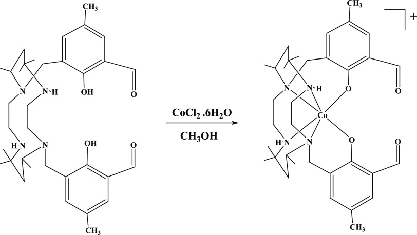
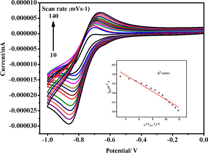
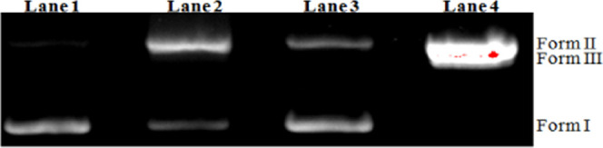
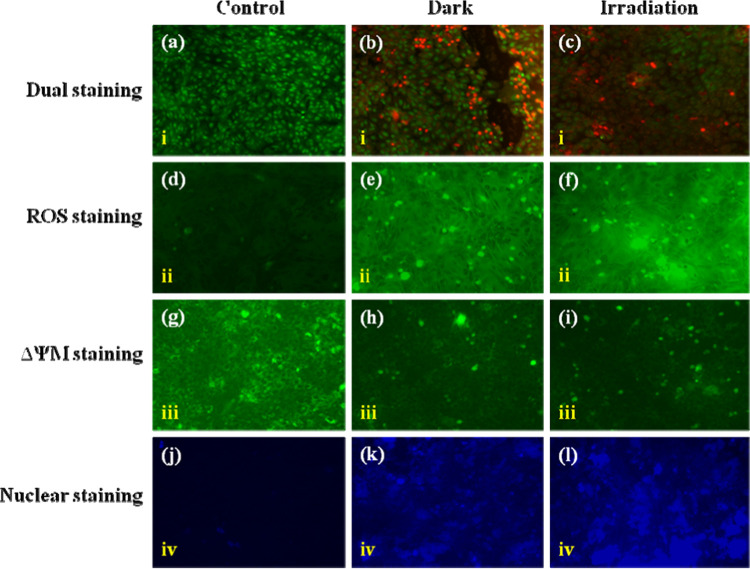
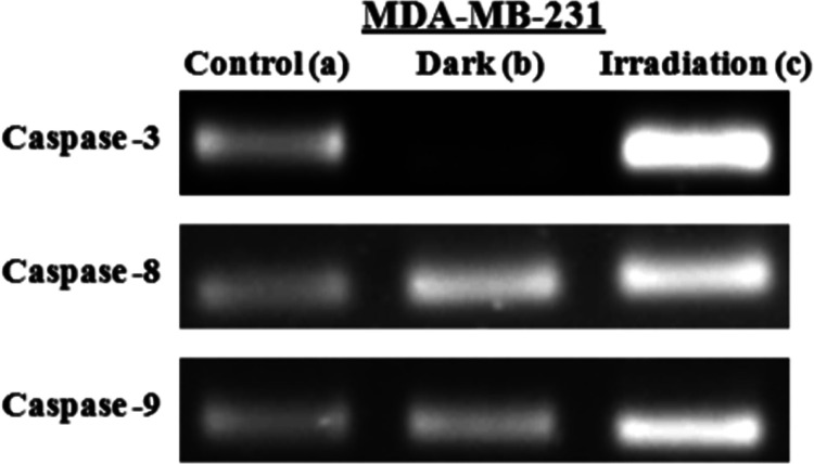
A cobalt(III) complex, [Co(L)]Cl (complex , where L = 1,8-[,-bis{(3-formyl-2-hydroxy-5-methyl)benzyl}]-1,4,8,11-tetraaza-5,5,7,12,12,14-hexamethylcyclotetradecane) with distorted octahedral geometry has been synthesized and characterized using various spectroscopic techniques. The structure of the ligand has remarkably rich hydrogen intermolecular interactions such as H···H, H···C/C···H, and H···O/O···H that vary with the presence of the metal ion, and the structure of complex has Cl···H interactions; this result has been proved by Hirshfeld surface and two-dimensional (2D) fingerprint maps analyses. The complex exhibits a quasi-reversible Co(III)/Co(II) redox couple with = -0.76 V. Calf thymus DNA (CT DNA) binding abilities of the ligand and complex were confirmed by spectroscopic and electrochemical analyses. According to absorption studies, the ligand and complex bind to CT DNA via intercalative binding mode, with intrinsic binding strengths of 1.41 × 10 and 8.64 × 10 M, respectively. A gel electrophoresis assay shows that complex promotes the pUC19 DNA cleavage under dark and light irradiation conditions. Complex has superior antimicrobial activity than the ligand. The cytotoxicity of complex was tested against MDA-MB-231 breast cancer cells with values of IC of 1.369 μg mL in the dark and 0.9034 μg mL after light irradiation. Besides, cell morphological studies confirmed the morphological changes with AO/EB dual staining, reactive oxygen species (ROS) staining, mitochondria staining, and Hoechst staining on MDA-MB-231 cancer cells by fluorescence microscopy. Complex was found to be a potent antiproliferative agent against MDA-MB-231 cells, and it can induce mitochondrial-mediated and caspase-dependent apoptosis with activation of downregulated caspases. The biotoxicity assay of complex on the development of was evaluated at an IC value of 200 μg mL and with excellent biocompatibility.
合成了一种具有扭曲八面体几何结构的钴(III)配合物[Co(L)]Cl(配合物 ,其中L = 1,8-[,-双{(3-甲酰基-2-羟基-5-甲基)苄基}]-1,4,8,11-四氮杂-5,5,7,12,12,14-六甲基环十四烷),并使用各种光谱技术对其进行了表征。配体的结构具有非常丰富的分子间氢键相互作用,如H···H、H···C/C···H和H···O/O···H,这些相互作用会随着金属离子的存在而变化,配合物 的结构具有Cl···H相互作用;这一结果已通过Hirshfeld表面和二维(2D)指纹图谱分析得到证实。该配合物表现出准可逆的Co(III)/Co(II)氧化还原对, = -0.76 V。通过光谱和电化学分析证实了配体和配合物 与小牛胸腺DNA(CT DNA)的结合能力。根据吸收研究,配体和配合物 通过插入结合模式与CT DNA结合,内在结合强度分别为1.41×10和8.64×10 M。凝胶电泳分析表明,配合物 在黑暗和光照条件下均能促进pUC19 DNA的切割。配合物 比配体具有更强的抗菌活性。测试了配合物 对MDA-MB-231乳腺癌细胞的细胞毒性,黑暗中IC值为1.369 μg mL,光照后为0.9034 μg mL。此外,细胞形态学研究通过荧光显微镜对MDA-MB-231癌细胞进行AO/EB双重染色、活性氧(ROS)染色、线粒体染色和Hoechst染色,证实了细胞形态的变化。发现配合物 是一种针对MDA-MB-231细胞的有效抗增殖剂,它可以通过激活下调的半胱天冬酶诱导线粒体介导的和半胱天冬酶依赖性凋亡。在IC值为200 μg mL时评估了配合物 对 的生物毒性,并发现其具有优异的生物相容性。