Department of Molecular and Developmental Medicine, Surgical and Neurological Sciences, University of Siena, Siena, Italy.
Department of Clinical, Surgical and Neurological Sciences, University of Siena, Siena, Italy.
Lymphat Res Biol. 2022 Oct;20(5):468-477. doi: 10.1089/lrb.2021.0090. Epub 2022 Jan 17.
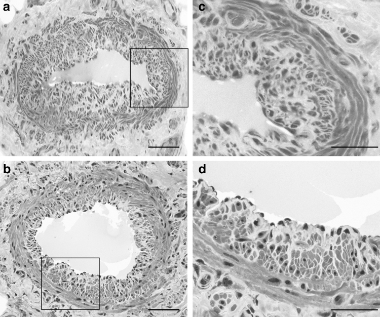
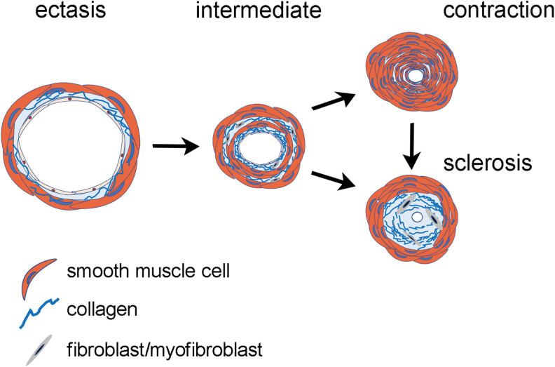
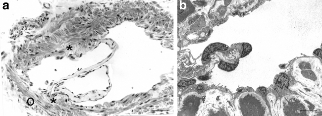
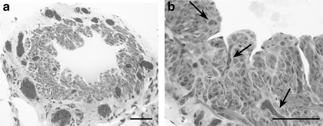
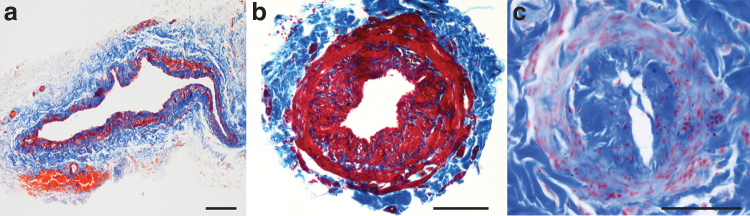
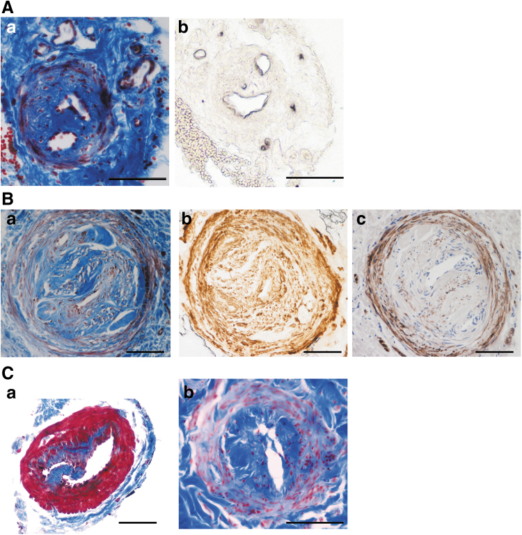
Secondary lymphedema of the extremities affects millions of people in the world as a common side effect of oncological treatments with heavy impact on every day life of patients and on the health care system. One of the surgical techniques for lymphedema treatment is the creation of a local connection between lymphatic vessels and veins, facilitating drainage of lymphatic fluid into the circulatory system. Successful results, however, rely on using a functional vessel for the anastomosis, and vessel function, in turn, depends on its structure. The structure of lymphatic collecting vessels changes with the progression of lymphedema. They appear initially dilated by excess interstitial fluid entered at capillary level. The number of lymphatic smooth muscle cells in their media then increases in the attempt to overcome the impaired drainage. When lymphatic muscle cells hyperplasia occurs at the expenses of the lumen, vessel patency decreases hampering lymph flow. Finally, collagen fiber accumulation leads to complete occlusion of the lumen rendering the vessel unfit to conduct lymph. Different types of vessels may coexist in the same patient but usually the distal part of the limb contains less affected vessels that are more likely to perform efficient lymphatic-venular anastomosis. Here we review the structure of the lymphatic collecting vessels in health and in lymphedema, focusing on the histopathological changes of the lymphatic vessel wall based on the observations on segments of the vessels used for lymphatic-venular anastomoses.
继发性肢体淋巴水肿是一种常见的肿瘤治疗的副作用,影响着全球数以百万计的人,对患者的日常生活和医疗保健系统都有重大影响。淋巴水肿治疗的手术技术之一是在淋巴管和静脉之间建立局部连接,促进淋巴液排入循环系统。然而,成功的结果依赖于吻合术使用功能血管,而血管功能又取决于其结构。淋巴收集血管的结构随着淋巴水肿的进展而改变。它们最初由于毛细血管水平进入的过多间质液而扩张。然后,为了克服受损的引流,其中的淋巴平滑肌细胞数量增加。当淋巴平滑肌细胞增生以牺牲管腔为代价时,血管通畅性降低,阻碍淋巴流动。最后,胶原纤维积累导致管腔完全闭塞,使血管无法输送淋巴。不同类型的血管可能存在于同一患者中,但通常肢体的远端部分包含较少受影响的血管,这些血管更有可能进行有效的淋巴静脉吻合术。在这里,我们回顾了健康和淋巴水肿状态下的淋巴收集血管的结构,重点关注基于用于淋巴静脉吻合术的血管段的观察,探讨了淋巴血管壁的组织病理学变化。