Department of Plastic and Reconstructive Surgery, The University of Tokyo, Tokyo, Japan.
PLoS One. 2012;7(7):e41126. doi: 10.1371/journal.pone.0041126. Epub 2012 Jul 24.
To date, an electron microscopy study of the collecting lymphatic vessels has not been conducted to examine the early stages of lymphedema. However, such histological studies could be useful for elucidating the mechanism of lymphedema onset. The aim of this study was to clarify the changes occurring in collecting lymphatic vessels after lymphadenectomy.
The study was conducted on 114 specimens from 37 patients who developed lymphedema of the lower limbs after receiving surgical treatment for gynecologic cancers and who consulted the University of Tokyo Hospital and affiliated hospitals from April 2009 to March 2011. Lymphatic vessels that were not needed for lymphatico venous anastomosis surgery were trimmed and subsequently examined using electron microscopy and light microscopy.
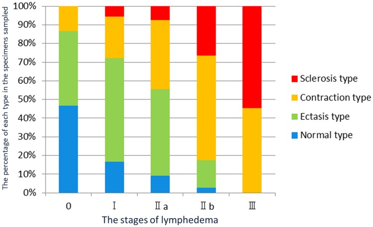
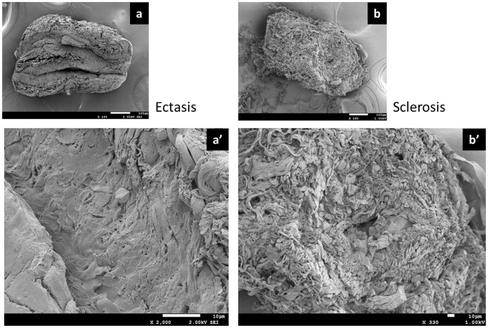
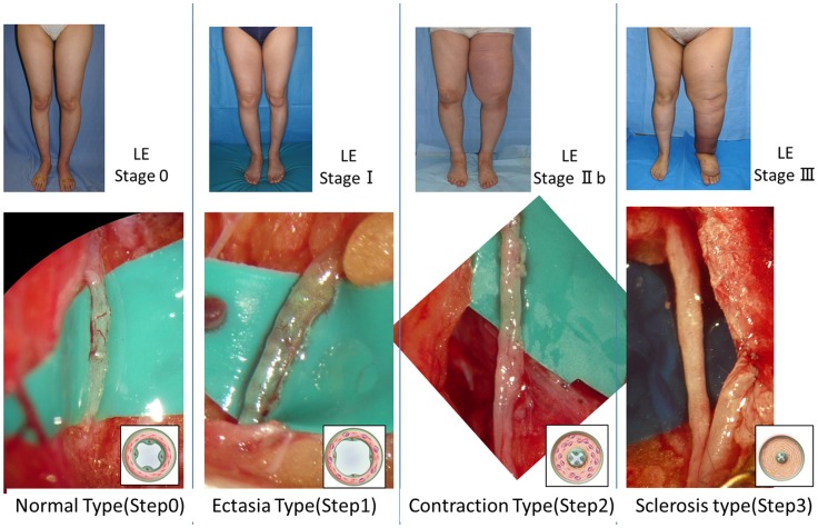
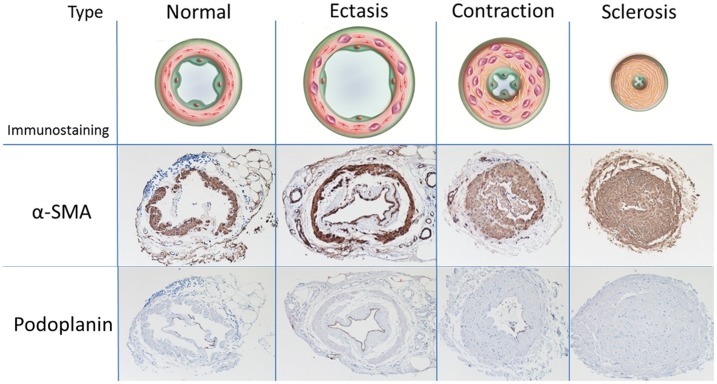
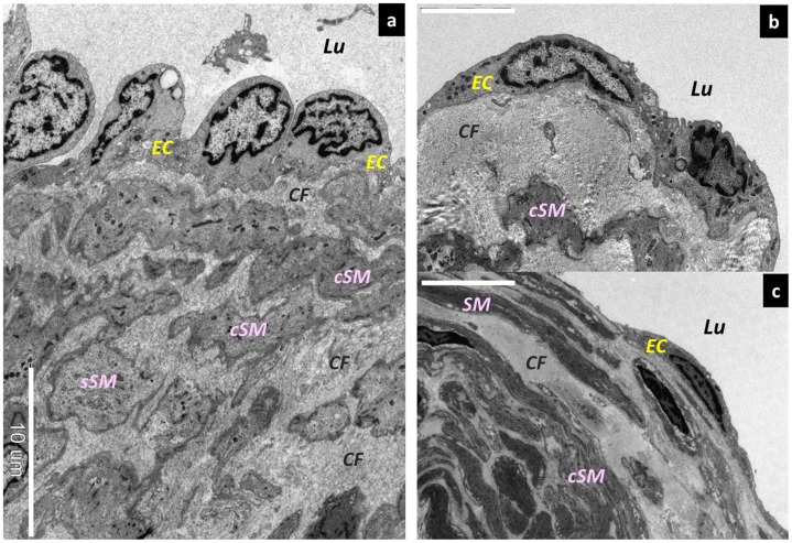
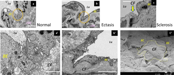
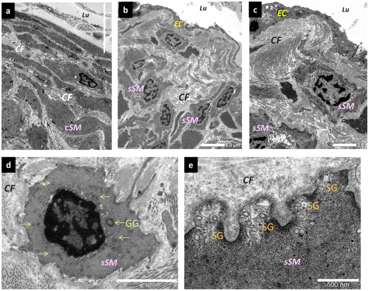
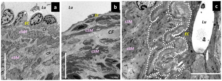
Based on macroscopic findings, the histochemical changes in the collecting lymphatic vessels were defined as follows: normal, ectasis, contraction, and sclerosis type (NECST). In the ectasis type, an increase in endolymphatic pressure was accompanied by a flattening of the lymphatic vessel endothelial cells. In the contraction type, smooth muscle cells were transformed into synthetic cells and promoted the growth of collagen fibers. In the sclerosis type, fibrous elements accounted for the majority of the components, the lymphatic vessels lost their transport and concentrating abilities, and the lumen was either narrowed or completely obstructed.
The increase in pressure inside the collecting lymphatic vessels after lymphadenectomy was accompanied by histological changes that began before the onset of lymphedema.
迄今为止,尚未对收集淋巴管进行电子显微镜研究,以检查淋巴水肿的早期阶段。然而,此类组织学研究对于阐明淋巴水肿发病机制可能是有用的。本研究旨在阐明淋巴结清扫术后收集淋巴管发生的变化。
该研究共纳入 37 例因妇科癌症接受手术治疗后出现下肢淋巴水肿并于 2009 年 4 月至 2011 年 3 月期间咨询东京大学医院及其附属医院的患者的 114 个标本。修剪不需要用于淋巴管静脉吻合术的淋巴管,然后使用电子显微镜和光镜进行检查。
根据宏观发现,将收集淋巴管的组织化学变化定义为:正常、扩张、收缩和硬化型(NECST)。在扩张型中,内淋巴压增加伴随着淋巴管内皮细胞变平。在收缩型中,平滑肌细胞转化为合成细胞并促进胶原纤维的生长。在硬化型中,纤维元素占大部分成分,淋巴管失去了其转运和浓缩能力,管腔变窄或完全阻塞。
淋巴结清扫术后收集淋巴管内压力增加伴随着淋巴水肿发病前开始的组织学变化。