Zhang Xun, Suo Xueling, Yang Xun, Lai Han, Pan Nanfang, He Min, Li Qingyuan, Kuang Weihong, Wang Song, Gong Qiyong
Huaxi MR Research Center (HMRRC), Department of Radiology, West China Hospital of Sichuan University, Chengdu, 610041, China.
Research Unit of Psychoradiology, Chinese Academy of Medical Sciences, Chengdu, 610041, China.
Transl Psychiatry. 2022 Jan 21;12(1):26. doi: 10.1038/s41398-022-01791-7.
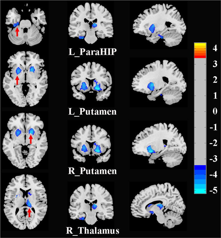
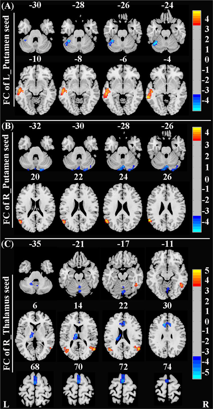
Although functional and structural abnormalities in brain regions involved in the neurobiology of fear and anxiety have been observed in patients with social anxiety disorder (SAD), the findings have been heterogeneous due to small sample sizes, demographic confounders, and methodological differences. Besides, multimodal neuroimaging studies on structural-functional deficits and couplings are rather scarce. Herein, we aimed to explore functional network anomalies in brain regions with structural deficits and the effects of structure-function couplings on the SAD diagnosis. High-resolution structural magnetic resonance imaging (MRI) and resting-state functional MRI images were obtained from 49 non-comorbid patients with SAD and 53 demography-matched healthy controls. Whole-brain voxel-based morphometry analysis was conducted to investigate structural alterations, which were subsequently used as seeds for the resting-state functional connectivity analysis. In addition, correlation and mediation analyses were performed to probe the potential roles of structural-functional deficits in SAD diagnosis. SAD patients had significant gray matter volume reductions in the bilateral putamen, right thalamus, and left parahippocampus. Besides, patients with SAD demonstrated widespread resting-state dysconnectivity in cortico-striato-thalamo-cerebellar circuitry. Moreover, dysconnectivity of the putamen with the cerebellum and the right thalamus with the middle temporal gyrus/supplementary motor area partially mediated the effects of putamen/thalamus atrophy on the SAD diagnosis. Our findings provide preliminary evidence for the involvement of structural and functional deficits in cortico-striato-thalamo-cerebellar circuitry in SAD, and may contribute to clarifying the underlying mechanisms of structure-function couplings for SAD. Therefore, they could offer insights into the neurobiological substrates of SAD.
尽管在社交焦虑障碍(SAD)患者中已观察到参与恐惧和焦虑神经生物学的脑区存在功能和结构异常,但由于样本量小、人口统计学混杂因素和方法学差异,研究结果一直存在异质性。此外,关于结构 - 功能缺陷及耦合的多模态神经影像学研究相当匮乏。在此,我们旨在探索存在结构缺陷的脑区中的功能网络异常以及结构 - 功能耦合对SAD诊断的影响。从49例无共病的SAD患者和53名人口统计学匹配的健康对照者获取了高分辨率结构磁共振成像(MRI)和静息态功能MRI图像。进行全脑基于体素的形态学分析以研究结构改变,随后将其用作静息态功能连接分析的种子点。此外,进行了相关性和中介分析以探究结构 - 功能缺陷在SAD诊断中的潜在作用。SAD患者双侧壳核、右侧丘脑和左侧海马旁回灰质体积显著减少。此外,SAD患者在皮质 - 纹状体 - 丘脑 - 小脑回路中表现出广泛的静息态连接障碍。而且,壳核与小脑以及右侧丘脑与颞中回/辅助运动区之间的连接障碍部分介导了壳核/丘脑萎缩对SAD诊断的影响。我们的研究结果为皮质 - 纹状体 - 丘脑 - 小脑回路中的结构和功能缺陷参与SAD提供了初步证据,并可能有助于阐明SAD结构 - 功能耦合的潜在机制。因此,它们可为SAD的神经生物学基础提供见解。