de Almeida Gonçalo Borges, Januário Gonçalo
Department of Neuroradiology, Hospital de São José, 1150-199 Lisbon, Portugal.
Department of Neurosurgery, Hospital de São José, 1150-199 Lisbon, Portugal.
Radiol Case Rep. 2022 Jan 15;17(3):922-927. doi: 10.1016/j.radcr.2021.12.059. eCollection 2022 Mar.
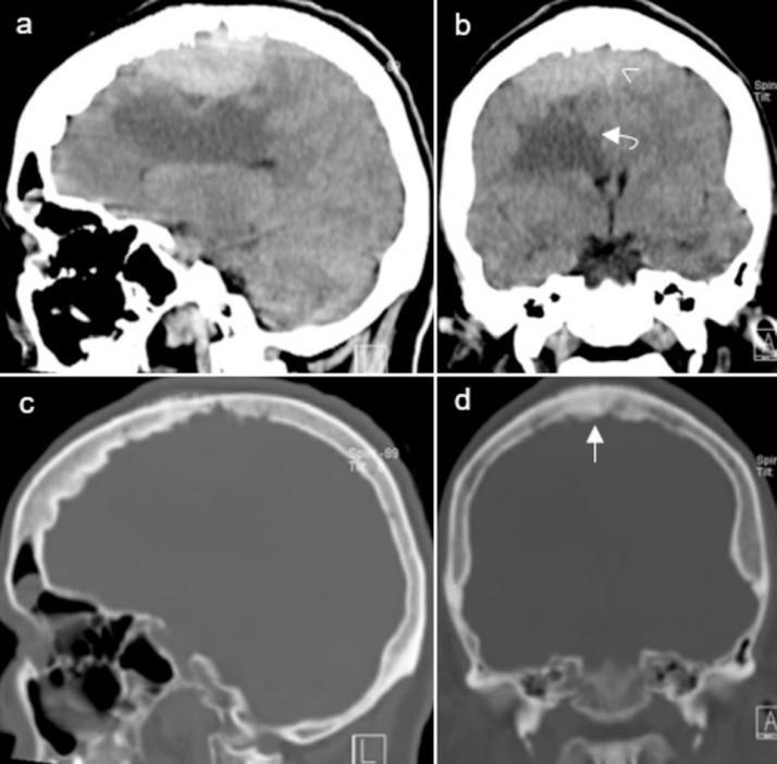
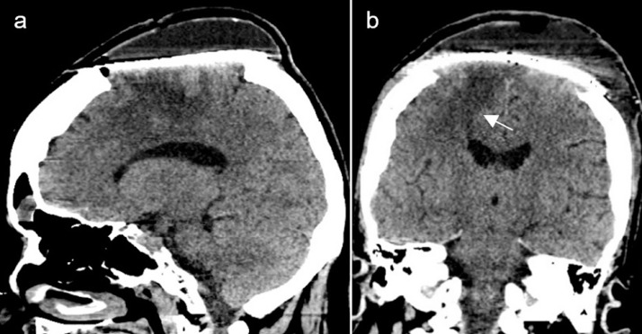
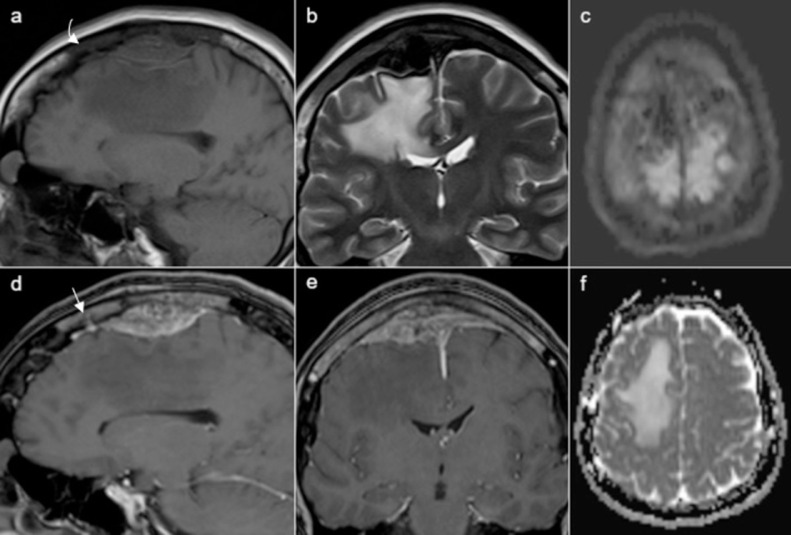
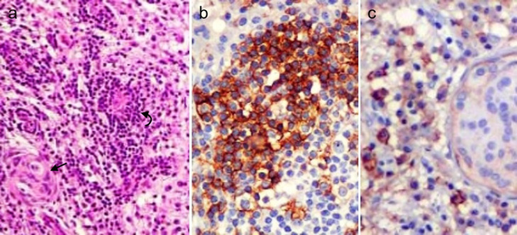
Meningiomas are the most common primary intracranial brain tumor and have been divided into 15 histologic subtypes, which are further classified into 3 grades according to biological behavior. Lymphoplasmacyte-rich meningioma is a rare histologic subtype of benign (grade 1) meningiomas characterized by prominent infiltration of plasma cells and lymphocytes, with a variable proportion of meningothelial elements. These benign meningioma variants usually cause significant peritumoral brain edema and mimic higher-grade lesions, which is believed to represent inflammatory cell infiltration rather than true neoplastic invasion. Bone invasion in these tumors is exceedingly rare and its clinical significance remains elusive. We describe the case of a lymphoplasmacyte-rich meningioma with skull invasion and peritumoral brain edema in a 57-year-old female patient presenting with left hemiparesis. Gross total resection of the lesion and adjacent skull were performed and histophatological examination disclosed a lymphoplasmacyte-rich meningioma. Gradual decrease of the parenchymal edema was seen on postoperative imaging studies and the patient showed progressive improvement of the motor deficit. This case report depicts rare bone invasion by lymphoplasmacyte-rich meningioma and highlights the other imaging features of this rare histologic subtype of benign meningioma. Due to the paucity of cases, gross total resection, and long-term follow-up are warranted as the prognosis of these tumors is still not fully understood.
脑膜瘤是最常见的原发性颅内脑肿瘤,已被分为15种组织学亚型,并根据生物学行为进一步分为3级。富于淋巴细胞浆细胞型脑膜瘤是良性(1级)脑膜瘤的一种罕见组织学亚型,其特征是浆细胞和淋巴细胞显著浸润,脑膜上皮成分比例不一。这些良性脑膜瘤变体通常会导致明显的瘤周脑水肿,并类似高级别病变,这被认为是炎症细胞浸润而非真正的肿瘤侵袭。这些肿瘤中的骨侵犯极为罕见,其临床意义仍不明确。我们描述了一例57岁女性患者,患有富于淋巴细胞浆细胞型脑膜瘤伴颅骨侵犯和瘤周脑水肿,表现为左半身轻瘫。对病变及相邻颅骨进行了全切术,组织病理学检查显示为富于淋巴细胞浆细胞型脑膜瘤。术后影像学检查显示实质水肿逐渐减轻,患者运动功能障碍逐渐改善。本病例报告描述了富于淋巴细胞浆细胞型脑膜瘤罕见的骨侵犯情况,并突出了这种罕见的良性脑膜瘤组织学亚型的其他影像学特征。由于病例稀少,鉴于这些肿瘤的预后仍未完全明确,进行全切术和长期随访是必要的。