Yan Yu, Balbastre Yaël, Brudfors Mikael, Ashburner John
Wellcome Centre for Human Neuroimaging, UCL Queen Square Institute of Neurology, University College London, London, United Kingdom.
School of Biomedical Engineering & Imaging Sciences, King's College London, London, United Kingdom.
Front Neurosci. 2022 Jan 17;15:818604. doi: 10.3389/fnins.2021.818604. eCollection 2021.
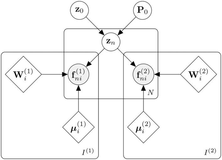
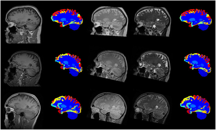
Segmentation of brain magnetic resonance images (MRI) into anatomical regions is a useful task in neuroimaging. Manual annotation is time consuming and expensive, so having a fully automated and general purpose brain segmentation algorithm is highly desirable. To this end, we propose a patched-based labell propagation approach based on a generative model with latent variables. Once trained, our Factorisation-based Image Labelling (FIL) model is able to label target images with a variety of image contrasts. We compare the effectiveness of our proposed model against the state-of-the-art using data from the . As our approach is intended to be general purpose, we also assess how well it can handle domain shift by labelling images of the same subjects acquired with different MR contrasts.
将脑磁共振成像(MRI)分割成解剖区域是神经成像中的一项有用任务。手动标注既耗时又昂贵,因此拥有一个完全自动化的通用脑分割算法是非常可取的。为此,我们提出了一种基于具有潜在变量的生成模型的基于补丁的标签传播方法。一旦训练完成,我们基于因子分解的图像标记(FIL)模型就能够用各种图像对比度对目标图像进行标记。我们使用来自[具体数据集未给出]的数据,将我们提出的模型与当前最先进的模型的有效性进行比较。由于我们的方法旨在具有通用性,我们还通过对用不同磁共振对比度采集的同一受试者的图像进行标记,来评估它处理域转移的能力。